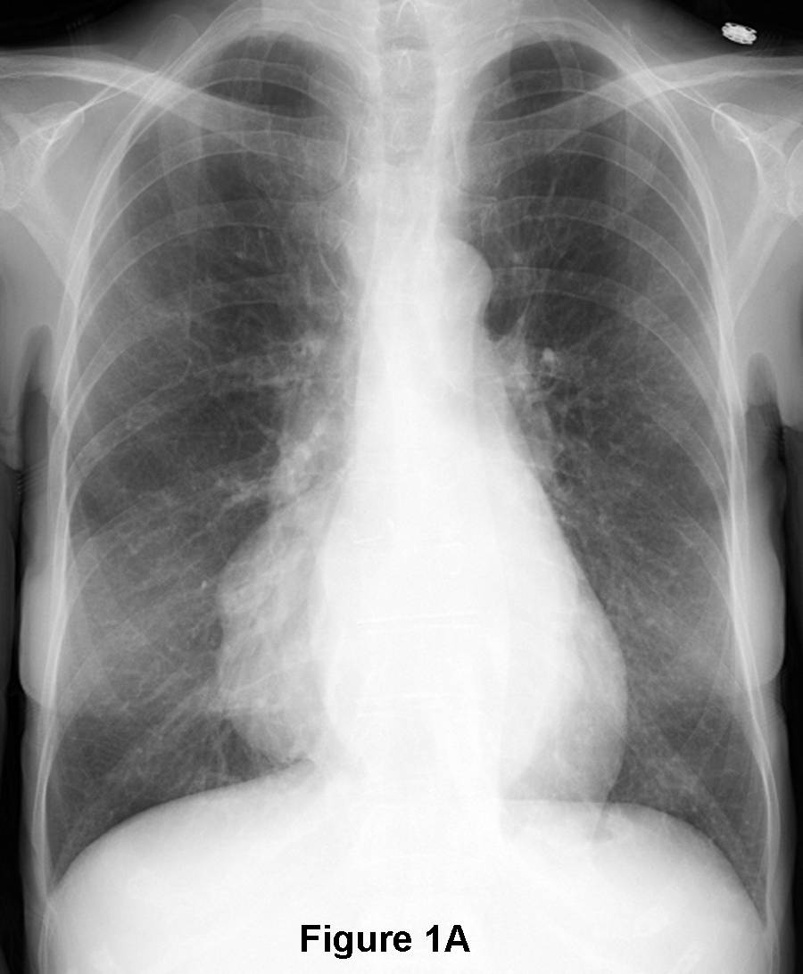
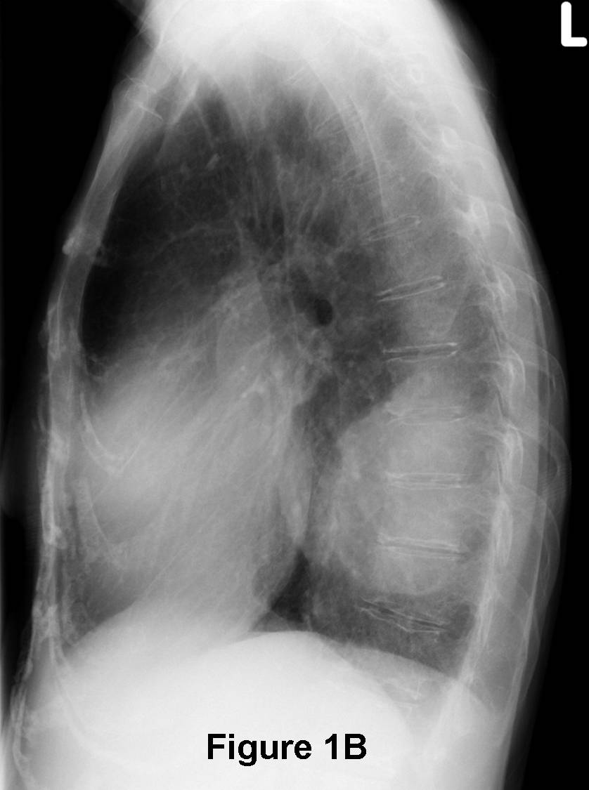
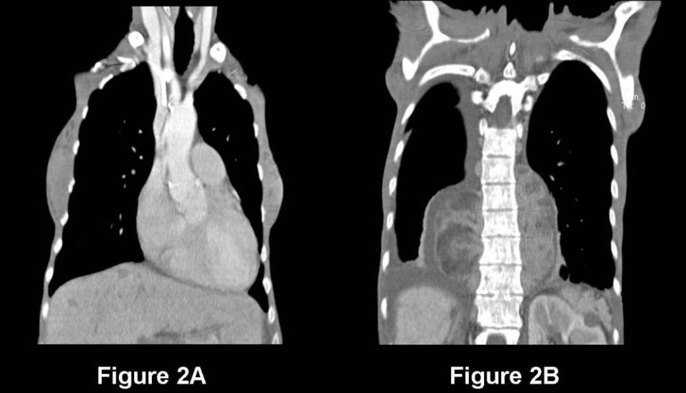
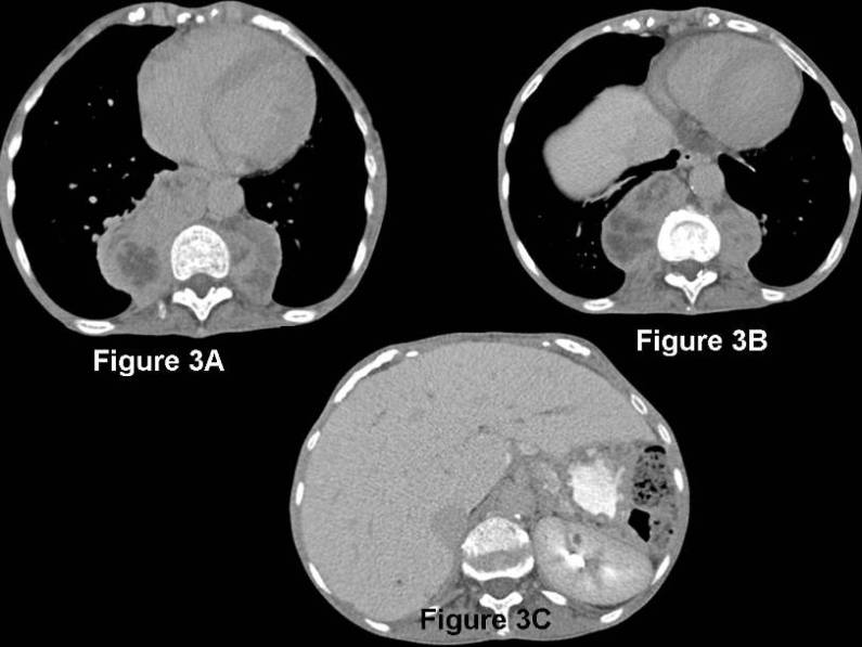
Radiologic Findings PA (Fig. 1A) and lateral (Fig. 1B) chest radiographs reveal bilateral, obtusely marginated, polylobular paramediastinal masses, right larger than left, that silhouette their respective paraspinal lines. That latter imaging finding localizes the masses to the posterior mediastinal paravertebral compartment. This is confirmed on the lateral exam (Fig. 1B). The masses are relatively homogenous in density and lack the presence of any calcifications. Contrast-enhanced coronal (Fig. 2A and 2B) and axial (Fig. 3A-3C) CT confirms the posterior mediastinal location of the masses. The masses are lobulated and contain intermingled regions of soft-tissue and fat attenuation. Note the expansile, permeative changes in the osseous structures from the marrow expansion and the hyperdense liver from previous transfusions and the surgical absence of the spleen. Diagnosis: Extramedullary Hematopoiesis
Differential Diagnosis
- Neurogenic Tumors
- Schwannoma
- Neurofibroma
- Malignant Peripheral Nerve Sheath Tumor
- Neuroblastic Tumors
- Neuroblastoma
- Ganglioneuroma
- Non-Neurogenic Tumors
- Chordoma
- Pheochromocytoma
- Paraspinal Abscess
- Descending Aorta Aneurysm
- Esophageal Neoplasia
- Hernias
- Lymphadenopathy / Lymphoma
- Foregut Duplication Cysts
- Neuroenteric Cysts
- Esophageal Duplication Cyst
- Thoracic Meningocele
Discussion
In the setting of a hemolytic or other chronic anemia, along with the imaging findings described above, extramedullary hematopoiesis should be the leading diagnosis. Extramedullary hematopoiesis (EMH) is the compensatory formation of blood elements outside of the osseous medulla, most commonly in the liver, spleen, and lymph nodes, but also in the posterior mediastinum. EMH usually occurs in response to inadequate erythrocyte production or excessive hemolysis. It is associated with severe congenital hemolytic anemias (e.g., hereditary spherocytosis, thalassemia major), polycythemia vera, lymphoproliferative disorders (e.g., lymphoma, chronic leukemia, myelofibrosis), and sickle cell anemia. It has been postulated that paravertebral extrusion of marrow elements through the abnormally thinned cortex of the adjacent ribs and vertebrae results in EMH.
Clinical Findings
EMH itself does not usually produce symptoms. Affected patients are usually diagnosed because of abnormal chest radiography acquired for unrelated reasons. There are rare reports of spinal cord compression and spontaneous hemothorax.
Imaging Findings
Chest Radiography
- Focal or multifocal smooth or polylobular paraspinal-posterior mediastinal masses
- Most commonly caudal to the 6th thoracic vertebra; may be located anywhere along the spine
- Sharply demarcated, lobulated, centered on vertebral bodies
- No vertebral body erosion
- No calcifications within the mass
- Very slow growth rate
CT - Well-defined polylobular soft-tissue mass; may contain fatty elements; the latter more commonly seen following splenectomy
- Marrow expansion of adjacent skeletal structures (e.g., lacy appearance or coarsened trabecullae)
- Will enhance heterogeneously with contrast
MRI
- Mass is higher in signal intensity than the adjacent marrow on T1WI / T2WI
Treatment
- Addressing the underlying anemia; providing sufficient transfusions if required
- In the case of spinal cord compression, EMH responds to small doses of radiation therapy
- Surgical resection may be attempted in severely symptomatic patients, frequently recurs
- Biopsy should be avoided if possible, as there is significant risk of bleeding.
Selected Readings
- Castelli R et al: Intrathoracic Masses due to Extramedullary Hematopoiesis. Am J Med Sci 2004; 328(5): 299-303.
- Edson et al. Extramedullary Hematopoiesis: Findings on Computed Tomography Scans of the Chest in 6 Patients. J Bras Pneumol [online] 2008; 34(10) [cited 11-20-2010], pp. 812-816.
- Parker MS, Rosado-de-Christenson ML, Abbott GF. Abnormalities and Diseases of the Mediastinum-Extramedullary Hematopoiesis. Teaching Atlas of Chest Imaging 2006; Thieme, New York, 695-698.
- Strollo DC, et. al, Primary Mediastinal Tumours, Part II. Tumors of the Middle and Posterior Mediastinum, Chest 1997; 112(5): 1344-1357.
Original case written by its authors at Virginia Commonwealth University and published at this address as part of a weekly teaching collection. Reproduced here as an archive.