Archived case 7 · Jul 9-Jul 16, 2009
Tracheobronchomegaly (Mounier-Kuhn Syndrome)
65-year-old man with a chronic productive cough and progressive dyspnea on exertion.
The question posed to readers
What is your diagnosis?
Archived case 7 · Jul 9-Jul 16, 2009
65-year-old man with a chronic productive cough and progressive dyspnea on exertion.
What is your diagnosis?
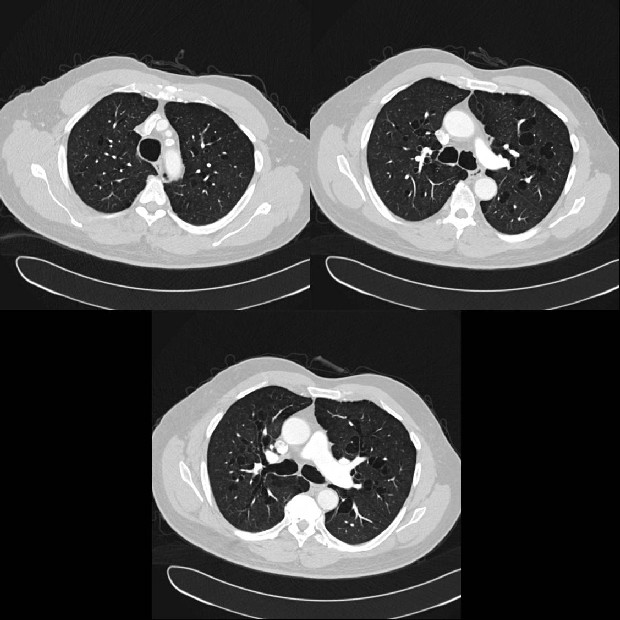
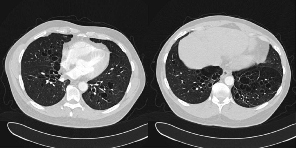
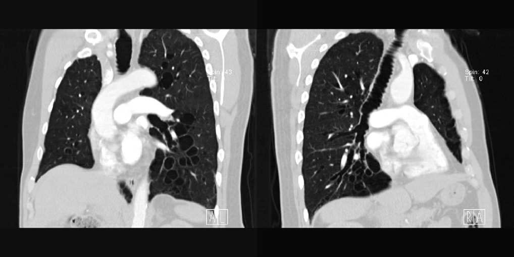
Tracheobronchomegaly (Mounier-Kuhn Syndrome)
Differential Diagnosis:
· Bronchiectasis
· Williams-Campbell Syndrome
o Normal trachea and central bronchi
o Dilatation of the 4th-6th order bronchi
· Complications of diffuse pulmonary fibrosis
· Chronic airway inflammation or infection with tracheobronchomalacia
· Allergic bronchopulmonary fungal disease including aspergillosis
Discussion:
Background
Tracheobronchomegaly or Mounier-Kuhn syndrome is a rare condition also known as tracheal diverticulosis and tracheobronchiectasis.
Etiology
The etiology of tracheobronchomegaly is unknown. Some authors have postulated a congenital etiology related to connective tissue disease because of associated conditions such as Ehlers-Danlos syndrome in affected adults and cutis laxa in affected children with reported associations of double carina, tracheal trifurcation, and a congenitally foreshortened right upper lobe bronchus. A familial form of the disease with autosomal-recessive inheritance has also been reported. Other authors postulate an acquired etiology, as the disease has been reported in infants after intensive ventilatory support. Cigarette smoking may also be implicated in the development of the disease. Tracheobronchomegaly is thought to result from weakness of cartilaginous and membranous components of the trachea and main bronchi.
Clinical Findings:
Affected patients are often men who are diagnosed in the third to fifth decades of life. Most affected adults present with recurrent pulmonary infection and marked sputum production, but symptoms usually date back to childhood. These patients may develop dyspnea on exertion and respiratory failure. Hemoptysis may occur. Spontaneous pneumothorax and digital clubbing have also been described. Some patients are entirely asymptomatic and are diagnosed incidentally.
Pathology:
Gross
· Dilatation of the trachea and main bronchi.
· Mucosal herniation between adjacent cartilage rings results in airway diverticulosis; diverticula may contain retained secretions.
· Normal caliber of fourth- and fifth-order airways.
Microscopic
· Atrophy and/or absence of longitudinal elastic fibers in the airway wall.
· Thinning of muscularis mucosa.
· Absent myenteric plexus.
· Absence of cartilaginous elements.
Imaging Findings:
Radiography
· Dilatation of trachea and main bronchi; may be limited to the trachea.
CT
· Dilatation of trachea and main bronchi.
· Diameter of trachea, right main bronchus or left main bronchus exceeding 30 mm, 24 mm, and 23 mm, respectively.
· Corrugated appearance of airway walls due to mucosal prolapse through adjacent cartilaginous rings.
· Perihilar cystic spaces representing bronchial diverticulosis.
· Proximal airway collapse on expiration due to tracheobronchomalacia.
Treatment:
· Postural drainage
· Bronchoscopy for clearance of secretions
· Antibiotics
· Tracheostomy and possible tracheal stenting
· Lung transplantation
Prognosis:
· Recurrent lower respiratory tract infections
· Development of dyspnea and respiratory failure as the lungs become progressively damaged
Caveats:
· Williams-Campbell syndrome results from absence of cartilage rings beyond the first and second bronchial divisions with resultant bronchiectasis that typically affects the fourth- to sixth-order bronchi. Affected patients have normal caliber trachea and central bronchi.
Suggested Readings:
1. Lazzarini-de-Oliveira LC, Costa de Barros Franco CA, Gomes de Salles CL, de Oliveira AC Jr. A 38-year-old man with tracheomegaly, tracheal diverticulosis, and bronchiectasis. Chest 2001; 120:1018-1020.
2. Maron EM, Goodman PC, McAdams HP. Diffuse abnormalities of the trachea and main bronchi. AJR Am J Roentgenol 2001; 176:713-717.
3. Parker MS, Rosado-de-Christenson ML, Abbott GF. Tracheobronchomegaly. Teaching Atlas of Chest Imaging. P.110-113.
4. Shin, et al. Tracheobronchomegaly (Mounier-Kuhn syndrome): CT diagnosis. AJR Am J Roentgenol. 1988 Apr; 150(4):777-779.
5. Woodring, et al. Congenital tracheobronchomegaly. (Mounier-Kuhn syndrome): a report of 10 cases and review of the literature. J Thorac Imaging 1991; 6(2):1-10.
Filed under: Radiology, Medicine/Pulmonary
Original case written by its authors at Virginia Commonwealth University and published at this address as part of a weekly teaching collection. Reproduced here as an archive.