Severe Right Ventricular Hypertrophy from Idiopathic Pulmonary Artery Hypertension
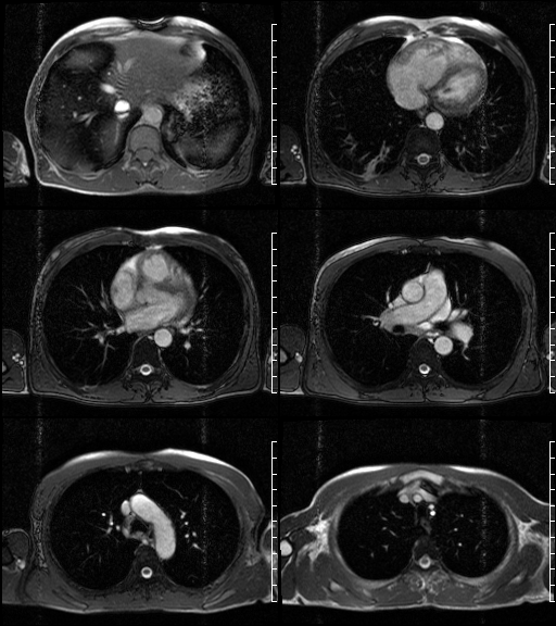
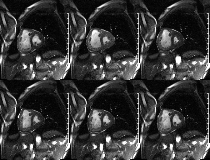
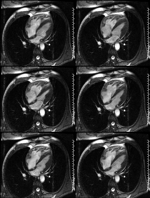
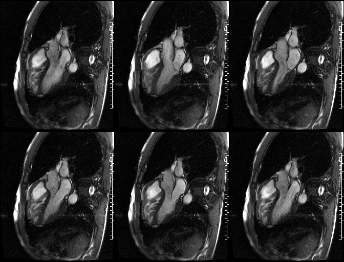
Selected images from cardiac MRI exam. Axial cine SSFP (Fig. 1) images demonstrate marked disproportionate enlargement of the right heart and right ventricle in particular as well as mild dilatation of the main pulmonary artery. No pericardial or pleural effusion is seen. Borderline sized lower right paratracheal and subcarinal lymph nodes and right lower lobe scarring is present. Short axis SSFP cine (Fig. 2) image shows not only right ventricular enlargement relative to the left ventricle, but marked right ventricular wall thickening with excessive hypertrophied trabecullae. Note the paradoxical septal bounce from the elevated pulmonary artery pressures (measured in excess of 60 mmHg on subsequent studies). The right ventricular ejection fraction is reduced (16%) and the right ventricle is globally hypokinetic. The left ventricle ejection fraction is normal (58%). 4-chamber SSFP cine (Fig. 3) image shows the hypertrophied right ventricular trabecullae thrown into redundant folds with the high signal blood pool extending into the interstices simulating right ventricular wall microaneurysms. Note the mild mitral regurgitation, right atrial enlargement, and moderate tricuspid regurgitation. No intracardiac septal defect is seen. 3-chamber SSFP cine (Fig. 4) image demonstrates many of the same imaging findings already addressed including the mild degree of mitral regurgitation. No aortic or pulmonary valvular insufficiency is seen. Delayed enhanced images revealed septal insertion site enhancement but no RV free wall enhancement (not illustrated). Diagnosis: Severe Right Ventricular Hypertrophy from Idiopathic Pulmonary Artery Hypertension
Differential Diagnosis Right Ventricular Hypertrophy
- Pulmonary Artery Hypertension
- Pulmonic Valvular Stenosis
- Intracardiac Septal Defect (VSD)
- Tetralogy of Fallot
- COPD
- Amyloidosis
- High-altitude
Discussion
Right ventricular hypertrophy (RVH) is commonly associated with any form of right ventricular outflow obstruction or pulmonary artery hypertension, which may in turn, may originate from left-sided disease. RVH is relatively rare. The four most common causes of RVH include: pulmonary artery hypertension (PAH); Tetralogy of Fallot (TOF); pulmonary valve stenosis (PVS); and ventricular septal defect (VSD). Marked hypertrophy of the right ventricular muscle can accentuate and exaggerate the normal trabecullae and its associated interstices and thereby mimic entities such as arrhythmogenic right ventricular dysplasia (ARVD) or cardiomyopathy (ARVCM). The latter is an inherited cardiomyopathy whose hallmark is right ventricular dilation and thinning (not hypertrophy); apical ventricular microaneurysms; and fibrofatty replacement of the right ventricular myocardium. Features simulated by but absent in this particular case. Additionally, delayed-hyperenhancement of the right ventricular free wall is often present in ARVD but not present in this case (not illustrated).
Clinical Findings
Mild RVH may be asymptomatic. Patients affected with more severe RVH may experience shortness of breath, chest pain, palpitations, near-syncope and syncope. Lower extremity swelling is not uncommon. ECG often reveals right-axis deviation. ARVD most commonly presents in young men (80% of cases in those under 40-years of age). Affected patients may likewise experience palpitations, syncope, and less commonly sudden cardiac death. The diagnostic criteria for ARVD are based on the presence of 2 major criteria or 1 major criterion plus 2 minor criteria or simply 4 minor criteria. Major criteria include: (1) structural abnormalities in the right ventricle; (2) abnormal right ventricle myocardial tissue with fatty infiltration; (3) T-wave inversion in leads V1 to V3 or beyond; (4) conduction abnormalities on the ECG; and (5) family history of ARVD confirmed by autopsy. Minor criteria include: (1) mild-to-moderate right ventricular structural abnormalities; (2) ECG abnormalities (e.g., T wave inversion in V2 and V3); (3) ventricular arrhythmias; (4) family history or premature sudden cardiac death or ARVD.
Selected Readings
- McLure LE, Peacock AJ. Cardiac magnetic resonance imaging for the assessment of the heart and pulmonary circulation in pulmonary hypertension. Eur Respir J. 2009; 33(6):1454-66. Review.
- Junqueira FP, Macedo R, Coutinho AC, et al. Myocardial delayed enhancement in patients with pulmonary hypertension and right ventricular failure: evaluation by cardiac MRI. Br J Radiol 2009; 82 (982): 821-826.
- Sen-Chowdhry S, Prasad SK, Syrris P, et al. Cardiovascular magnetic resonance arrhythmogenic right ventricular cardiomyopathy revisited: comparison with task force criteria and genotype. J Am Coll Cardiol. 2006; 48(10):2132-2140.
- Tandri H, et al. Noninvasive detection of myocardial fibrosis in arrhythmogenic right ventricular cardiomyopathy using delayed-enhancement magnetic resonance imaging. J Am Coll Cardiol. 2005; 45(1): 98-103.
Original case written by its authors at Virginia Commonwealth University and published at this address as part of a weekly teaching collection. Reproduced here as an archive.