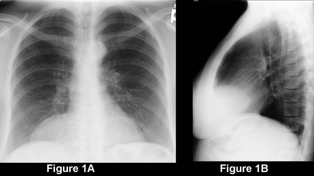
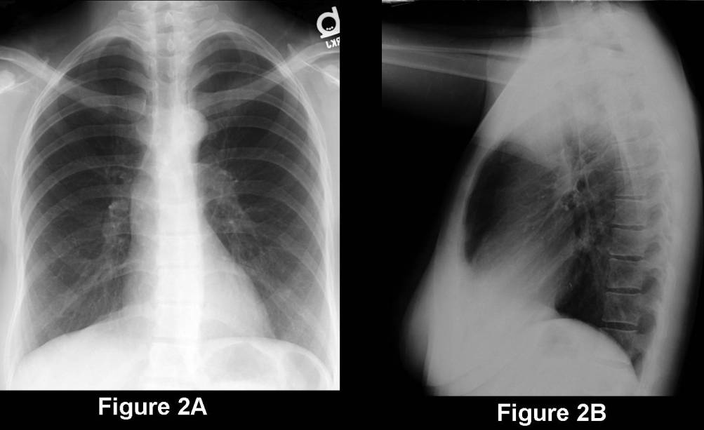
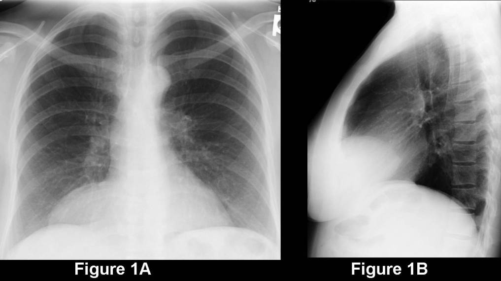
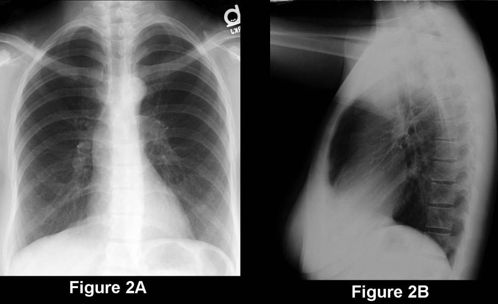
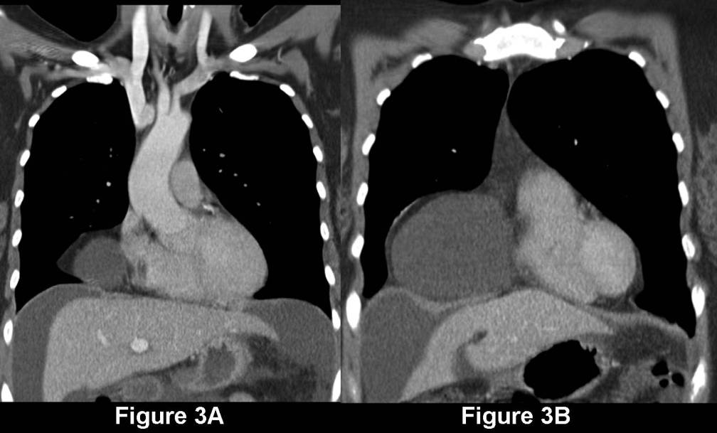
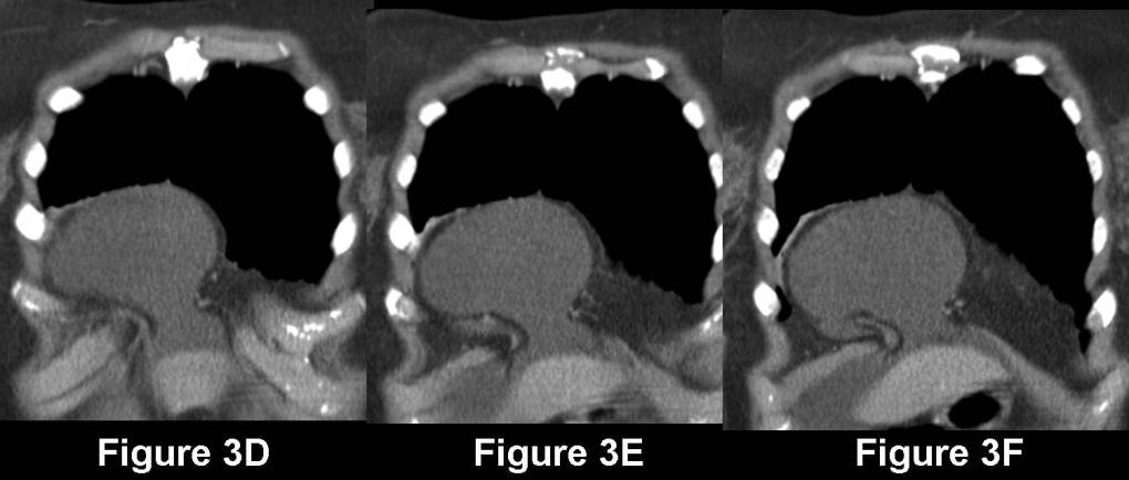
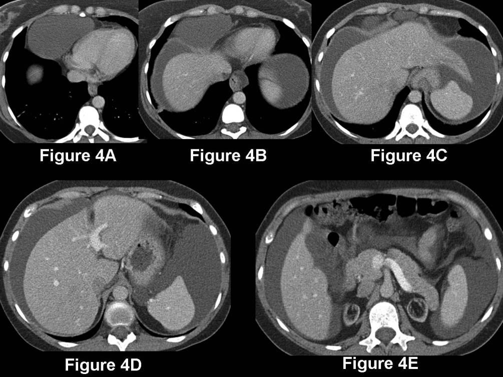
Baseline PA (Fig. 2A) and lateral (Fig. 2B) chest radiographs show well-expanded lungs and a normal appearing cardiomediastinal silhouette. The follow-up PA (Fig. 1A) and lateral (Fig. 1B) chest radiographs demonstrate interval development of a well-defined mass in the right anteromedial cardiophrenic angle and sulcus. The lesion does not appear to be very dense radiographically, as blood vessels can be perceived through the mass itself. This suggests the mass is composed of either fluid or fat. Contrast-enhanced coronal CT (Fig. 3A-3F) (mediastinal windows) confirms the cardiophrenic angle mass is indeed comprised on fluid. This is also a large volume of peritoneal fluid consistent with dialysate. Note the separation and diastasis of the anteromedial diaphragmatic leaves and extension of the peritoneal dialysate through the Morgagni-like defect into the cardiophrenic angle (Fig. 3AD-3F). Contrast-enhanced axial CT (Fig. 4A-4E) (mediastinal windows) confirm the above findings and also reveal marked atrophy of both kidneys related to the underlying chronic renal disease. Diagnosis: Morgagni-Type Defect with Herniation of Peritoneal Dialysate
Differential Diagnosis Cardiophrenic Angle Masses
- Abscess / Hydatid Disease
- Aneurysm (e.g., Right Atrium; Ventricle; Sinus of Valsalva)
- Cardiac Lesions
- Foregut Duplication Cysts
- Lipoma
- Lymphadenopathy (e.g., Tuberculosis; Hodgkin Lymphoma; Breast Cancer)
- Morgagni Hernia
- Neurogenic Tumors
- Paracardiac Varices
- Pericardial Lesions
- Pericardial Cyst
- Primary and Secondary Intrapericardiac Neoplasms
- Primary Lung Neoplasia
- Pronounced Pleural-Pericardial fat Pad
- Thymoma
- Thymolipoma
Differential Diagnosis Low-Density Cardiophrenic Angle Masses
- Foregut Duplication Cysts
- Lipoma
- Lymphadenopathy (e.g., Tuberculosis; Hodgkin Lymphoma; Breast Cancer)
- Morgagni Hernia
- Pericardial Cyst
- Pronounced Pleural-Pericardial fat Pad
- Thymolipoma
Discussion
There are 3 non traumatic hernias of the diaphragm. These include:
(1) Hiatus hernia- occurs at the esophageal hiatus. Sliding or paraesophageal varieties exist. On chest radiography, the hernia is often seen projecting posterior to the heart and an air/fluid level may be present. Esophography is confirmatory.
(2) Bochdalek- results from a defect in the embryonic pleuroperitoneal canal. Large hernias present in the neonatal period with hypoplasia of the ipsilateral lung. Small hernias may go undetected in childhood and are a common incidental finding in adults. Such hernias often appear as a posterolateral mass above the left hemidiaphragm, but may occur anywhere along the posterior diaphragmatic surface. MDCT nicely depicts the diaphragmatic defect and often reveals herniation of retroperitoneal fat, omentum, spleen, or kidney.
(3) Morgagni Hernia -results from a defect in the parasternal portion of the diaphragm, at the foramen of Morgagni, and is the least common of the three types of hernia. Morgagni hernias are more common in women and are rarely associated with other congenital abnormalities. These hernias are usually right-sided (90%) and appear as an asymptomatic cardiophrenic angle mass. The foramen of Morgagni is a small diaphragmatic cleft bound medially by muscle fibers originating from the sternum and laterally by the seventh intercostal cartilage. The diagnosis is made by noting herniation of omental fat, liver, transverse colon, or as in this case dialysate through the paracardiac portion of the right hemi-diaphragm. This is an unusual presentation of a Morgagni-type hernia, but when considering cardiophrenic angle masses, all aspects of patient history and symptomatology must be considered (i.e. current peritoneal dialysis)
Treatment
Adults:
- Surgical repair of Morgagni hernias:
- Usually only indicated when symptoms are present; some authors recommend surgical repair even in asymptomatic patients because of the risk of strangulation
- Open surgical repair via an abdominal, or transthoracic approach has traditionally been performed
- Regardless of the approach, the goals of the operation are the same: reduction of hernia contents, excision of hernia sac and closure of diaphragmatic defect to prevent recurrences
- Recent advances in minimally invasive surgery have led to reports of closure of foramen of Morgagni hernias using laparoscopic and thoracoscopic techniques
Selected Readings
- Brant, William E. and Helms, Clyde A. Fundamentals of Diagnostic Radiology; 2007.
- Michel, Steven J. and Bensadoun, Eric S. A Mass at the Right Cardiophrenic Angle. Respiration 2005; 72: 301-303
- Parker, Mark S., Rosado-de-Christenson, Melissa L., and Abbott, Gerald F. Teaching Atlas of Chest Imaging; 2006
- Pineda Victor, Andréu Jordi, Cáceres José, Merino Xavier, Varona Diego, Domínguez-Oronoz Rosa. Lesions of the Cardiophrenic Space: Findings on Cross-Sectional Imaging RadioGraphics January 2007; 19-32.
Original case written by its authors at Virginia Commonwealth University and published at this address as part of a weekly teaching collection. Reproduced here as an archive.