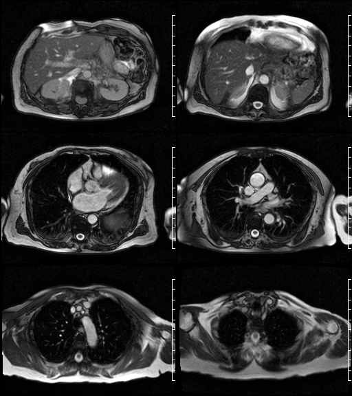
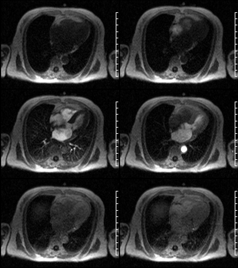
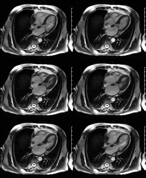
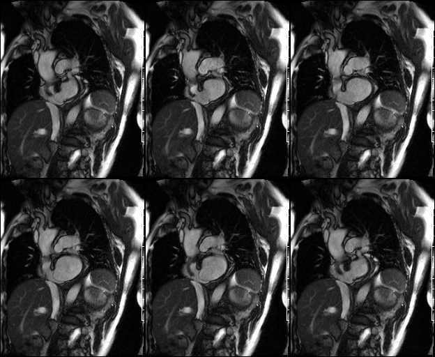
Selected images from a cardiac MRI. Axial FISP (Fig. 1); T1-axial perfusion (Fig. 2); 4-chamber cine FISP (Fig. 3); sagittal oblique cine FISP (Fig. 4) images demonstrate a redundant, thin, floppy, hypermobile interatrial septum that balloons at least 10 mm back and forth between the right and left atrium during the cardiac cycle. Perfusion imaging (Fig. 2) however does NOT demonstrate any flow through a patent foramen ovale (PFO) (Images courtesy of John D. Grizzard, MD, VCU Medical Center, Richmond, Virginia). Diagnosis: Atrial Septal Aneurysm; Type 5
Differential Diagnosis
None
Discussion
Atrial Septal Aneurysm (ASA) is a localized, saccular, deformity of the interatrial septum that usually occurs at the fossa ovalis and often bulges into the right atrium, left atrium, or both during the cardiac cycle. The prevalence of ASA varies, but is estimated between 0.08% and 1.2%. ASA may be “fixed” or bulge only within one atrium (46%), or may “mobile” and bulge “bi-directionally” into both atria during the cardiac cycle (54%). Alternatively, ASA be further classified into one of five types: Type 1R-ASA protrudes from the midline of the septum into the right atrium; Type 2L- ASA protrudes from the midline of the septum into the left atrium; Type 3RL-maximum excursion of the ASA is toward the right atrium, with a lesser excursion toward the left atrium; Type 4LR-maximum excursion of the ASA is toward the left atrium, with a lesser excursion toward the right atrium; and Type 5- ASA movement is “bidirectional” and “equidistant” between the right and left atrium during the cardiac cycle.
Clinical Findings
Atrial septal aneurysm may occur as an isolated finding in up to 32% of patients. However, ASA has also been associated with various congenital heart defects such as patent foramen ovale (PFO), atrial septal defects (ASD), ventricular septal defects (VSD), valvular prolapse (VP), patent ductus arteriosus (PDA), and Ebstein anomaly. 80-90% of patients with ASA will have a PFO. Embolism can result from thrombus formation within the aneurysm or occur as a paradoxical embolism. A PFO however was not demonstrated in this particular case. ASA has also been reported in association with various acquired heart diseases including valvular disease, cardiomyopathy, pulmonary artery hypertension, ischemic heart disease, and atrial dysrhythmias.
Imaging Findings
Echosonography or MRI
- Cutoff point between a redundant atrial septum and an ASA is somewhat arbitrary
- ASA is usually characterized by a thin, localized out-pouching of the middle portion of the atrial septum, but not involving the entire septum
- Protrusion of the aneurysm >10 mm beyond the plane of the atrial septum into either the right or left atrium
Caveats
Both atrial septal aneurysm and patent foramen ovale have been associated with an increased risk of cerebral vascular accident (CVA) and transient ischemic attacks (TIA’s).
Selected Readings
- Chidambaram M, Mink S, Sharma S. Atrial Septal Aneurysm with Right-to-Left Shunting. Tex Heart Inst J. 2003; 30(1): 68-70.
- Grizzard JD, Judd RM, Kim RJ. Teaching File Case 61. In; Cardiovascular MRI in Practice. Springer-Verlag, London, 2008; 163.
- Mügge A, Daniel WG, Angermann C, et al. Atrial Septal Aneurysm in Adult Patients: A Multicenter Study Using Transthoracic and Transesophageal Echocardiography Circulation. 1995; 91:2785-2792.
- Olivares-Reyes A; Al-Kamme A; Gonzalez J. Atrial Septal Aneurysm: A Study in Five Hundred Adult Patients. Premio al mejor Tema Libre Realizado por Médicos Residentes
Original case written by its authors at Virginia Commonwealth University and published at this address as part of a weekly teaching collection. Reproduced here as an archive.