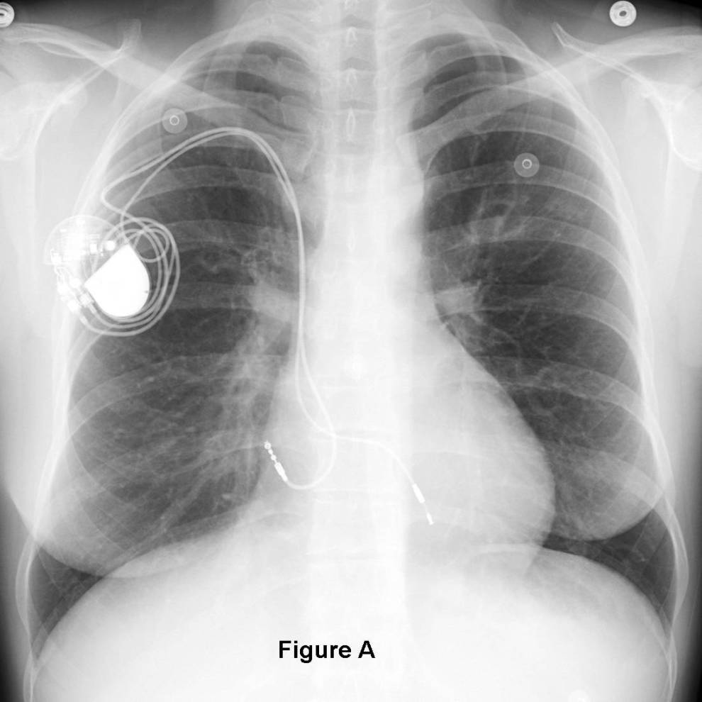
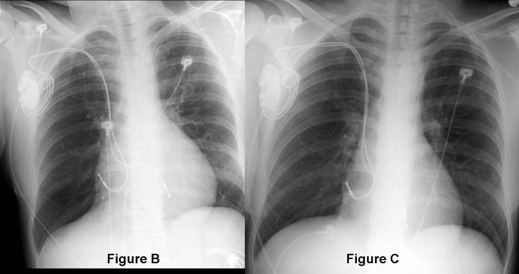
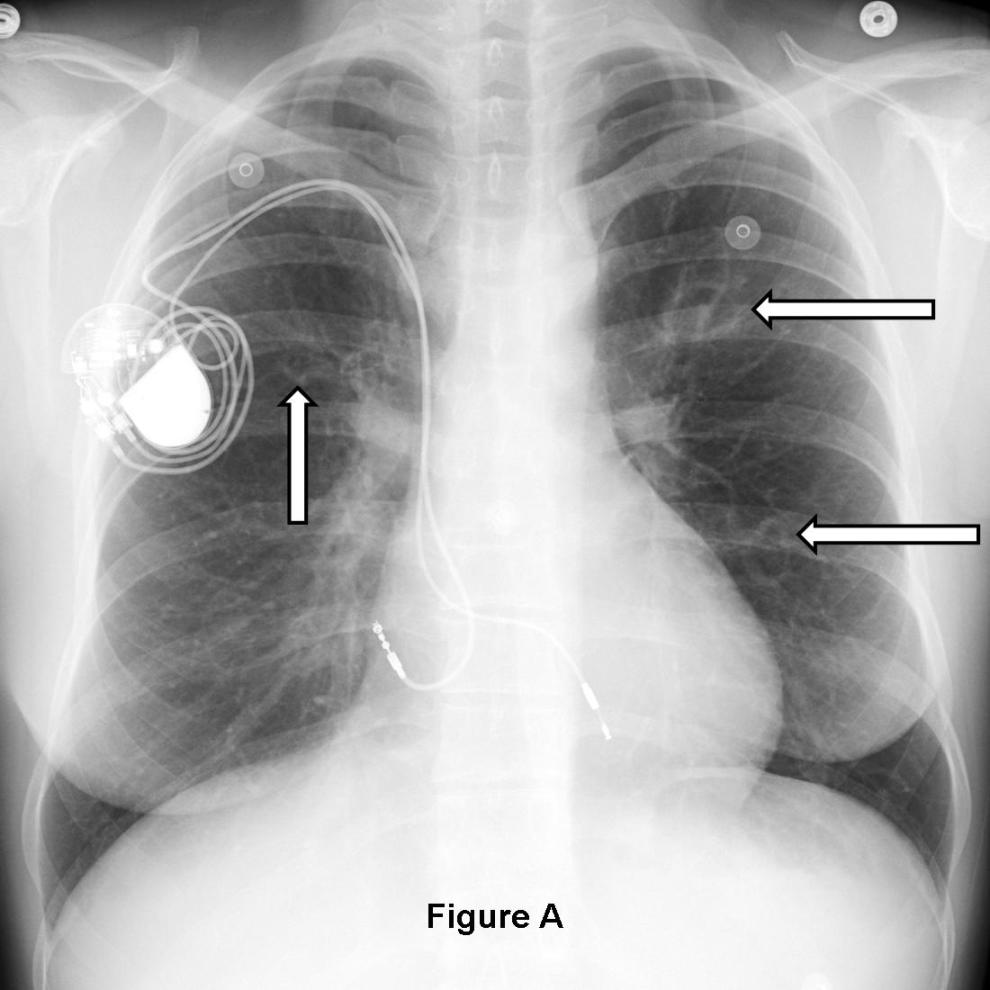
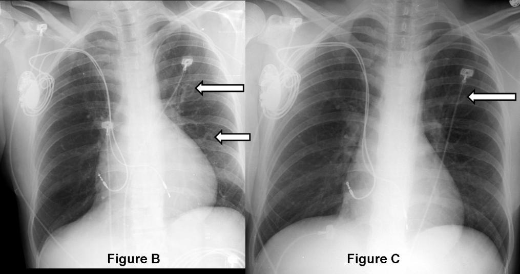
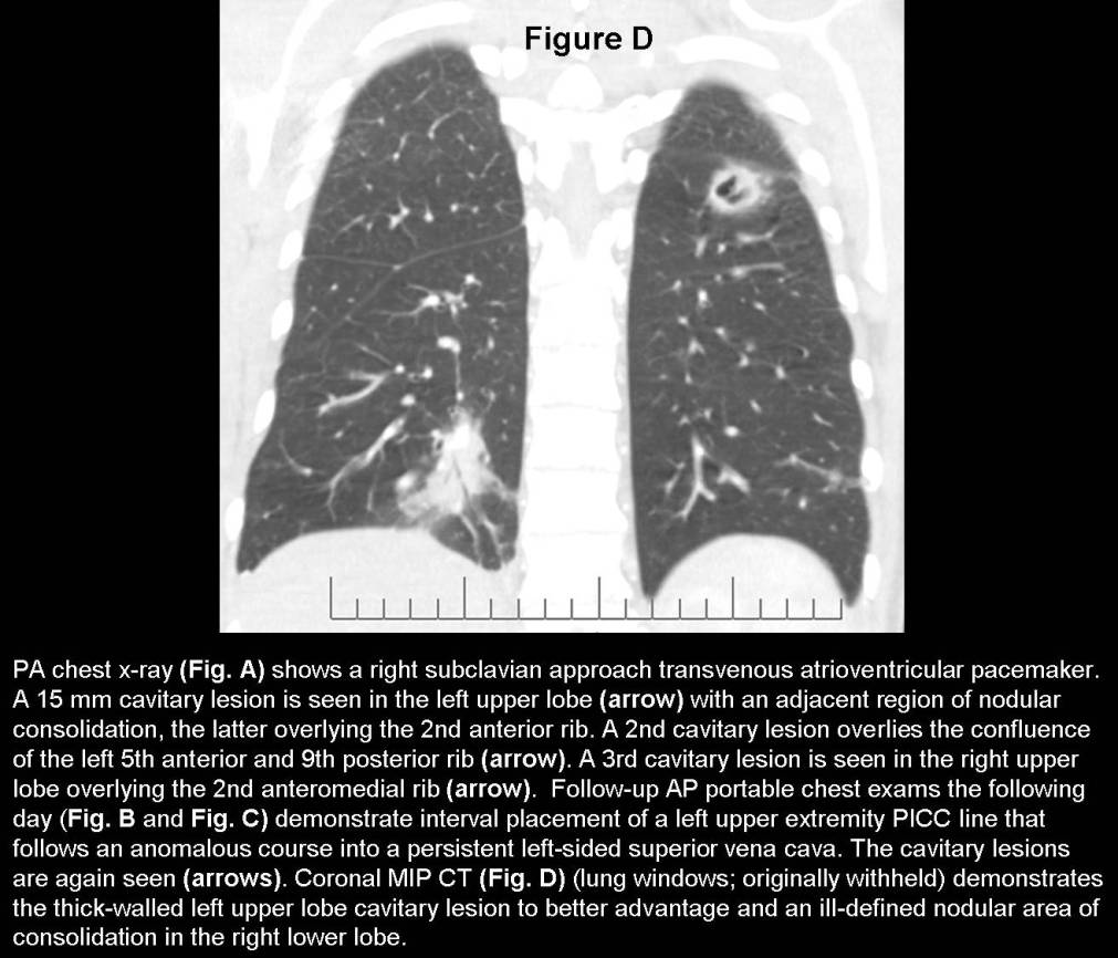
Young woman with remote history of congenital heart disease and septal defect repaired as a child. Currently, the patient has fever, bacteremia, intermittent hemoptysis, and endocarditis (history originally withheld). You are shown a series of chest x-rays. Diagnosis: Septic Pulmonary Emboli; Persistent Left-sided SVC
Differential Diagnosis: Cavitary lung lesions (Table-1)
Table-1: Differential Diagnosis Cavitary Lung Lesions
Infection Neoplasia Autoimmune Vascular Miscellaneous S. aureus Squamous Cell Wegener Granulomatosis Septic Emboli Traumatic Lung Cysts
S. pneumoniae Adenocarcinoma Systemic Lupus Erythematosis Pulmonary Emboli with Infarction Bronchiectasis
H. influenzae Transitional cell Bladder Rheumatoid Necrobiotic Nodules
K. pneumoniae Colorectal Cancer Blastomycosis Gynecologic Cancers Histoplasmosis Sarcomas Coccidioidomycosis Melanoma
Aspergillus sp. Germ Cell Tumors
M. tuberculosis Discussion
Persistent Left-sided Superior Vena Cava (SVC)
A persistent left SVC is a relatively common anomaly and represents the most frequent form of anomalous venous return to the heart. It occurs in approximately 0.3% of the general population, with an increased prevalence (4.4%) in patients with congenital heart disease. Embryologically, it results from failure of involution of the left anterior cardinal vein.
Cysts versus Cavities
Cysts represent thin-walled (< 3 mm in greatest thickness), well-defined and well-circumscribed air- or fluid-containing lesions. Common causes include bullae, blebs, pneumatoceles, various infections, and congenital lesions such as sequestration and Congenital Cystic Adenomatoid Malformation / CCAM (recently renamed Congenital Pulmonary Airway Malformation / CPAM) of the lung. Cavities represent lesions of varying size and wall thickness that likewise may contain air, fluid, and a combination of both. Cavities may be well-defined or ill-defined depending on the underlying etiology (Table-1).
Septic Emboli
Septic emboli most often originate from an extrapulmonary source that has become infected (e.g., intravenous access lines or ports, pacemakers and ICD devices, arteriovenous shunts, various grafts, prostheses, especially artificial cardiac valves, etc). Intravenous drug abuse (IVDA) with subsequent seeding of the cardiac valves and formation of valvular vegetation is also a not uncommon cause of septic emboli. Lemierre syndrome (head and neck thrombophlebitis is another potential source of septic embolic disease. Subsequent echosonography on this particular patient confirmed the diagnosis of tricuspid valvular vegetation and endocarditis. The most common bacterial organisms implicated and cultured in the setting of septic emboli include: Staphylococcus sp. Fusobacterium sp is also a not uncommon human pathogen. Aspergillus fumigatus has been associated with infected pacemaker leads. Salmonella has been associated with advanced HIV-AIDS infection.
Clinical Findings
Patients with septic pulmonary emboli often present with fever and shaking chills, cough, dyspnea, chest pain, and hemoptysis, the latter of which can be massive. Leukocytosis is common. Affected patients often have a history of IV drug abuse or the presence of indwelling prosthetic or mechanical devices.
Imaging Findings
Chest Radiography
- Variable size pulmonary nodules / masses; may or may not cavitate (Fig. A-C)
- Multi-focal rounded or wedge-shaped opacities
- Cavitary lesions when present may demonstrate varying degrees of wall thickness (Fig. A-C) and or air-fluid levels
- Lesions may be migratory; new lesions appear as pre-existing lesions fade
- Pleural effusion; uncommon
- Hilar and mediastinal lymphadenopathy; not uncommon
MDCT
- Variable number of primarily peripheral or juxtapleural lung nodules (Fig. D)
- Cavitary lesions when present may demonstrate varying degrees of wall thickness (Fig. D) and or air-fluid levels
- Foci of peripheral ground-glass or frank consolidation (Fig. D)
- Nodular opacities and area of consolidation often angiocentric (i.e., feeding vessel supplies the lesion)
Gated CTA / MR / Echosonography
- May reveal valvular vegetations
Treatment
- Antibiotics, often a prolonged course
- Infected prosthetic devices are removed if possible
Prognosis
Generally good with adequate and complete treatment
Selected Readings
- Cook RJ, Ashton RW, Aughenbaugh GL, Ryu JH. Septic pulmonary embolism: presenting features and clinical course of 14 patients. Chest 2005; 128(1):162-6. PubMed PMID: 16002930.
- Dodd JD, Souza CA, Müller NL. High-resolution MDCT of pulmonary septic embolism: evaluation of the feeding vessel sign. AJR Am J Roentgenol 2006; 187(3):623-629. PubMed PMID: 16928922.
- Gadkowski LB, Stout JE. Cavitary pulmonary disease. Clin Microbiol Rev 2008; 21(2):305-333. Review. PubMed PMID: 18400799.
- Parker MS, Rosado-de-Christenson ML, Abbott GF. StaphylococcalPneumonia. In: Teaching Atlas of Chest Imaging. Thieme, New York: 2006; 216-219.
- Ryu JH, Swensen SJ. Cystic and cavitary lung diseases: focal and diffuse. Mayo Clin Proc 2003; 78(6):744-52. Review. PubMed PMID: 12934786.
Original case written by its authors at Virginia Commonwealth University and published at this address as part of a weekly teaching collection. Reproduced here as an archive.