Archived case 55 · Jul 9-Jul 16, 2010
Total Artificial Heart (TAH) implantation
Clinical Presentation: Withheld
The question posed to readers
Please describe the surgical procedure that has been performed and its primary clinical indication
Archived case 55 · Jul 9-Jul 16, 2010
Clinical Presentation: Withheld
Please describe the surgical procedure that has been performed and its primary clinical indication
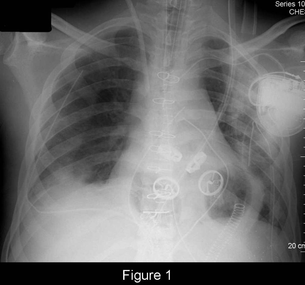
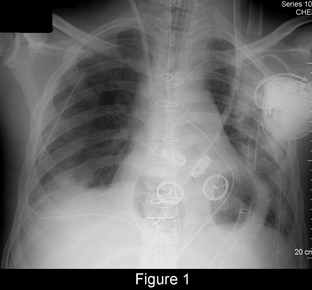
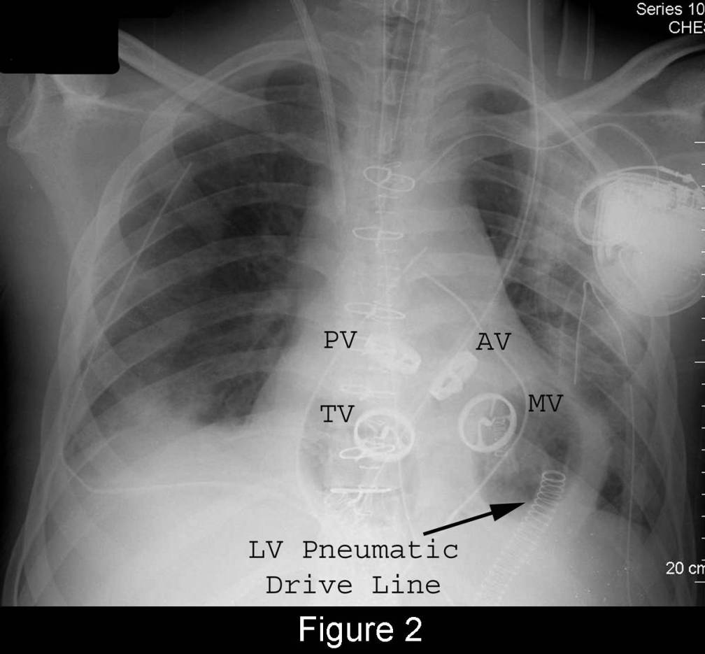
Total Artificial Heart (TAH) implantation
Differential Diagnosis
None
Discussion
The total artificial heart (TAH) is presently used in the United States as a bridge for patients with end-stage biventricular heart failure awaiting heart transplantation. This mechanical device is a biventricular orthotopic pneumatic pulsatile pump with two separate artificial ventricles that replace the patient’s native ventricles. Each artificial semi-rigid polyurethane ventricle contains a seamless blood-contacting diaphragm, two intermediate diaphragms, an air diaphragm, and an inflow and outflow mechanical valve. A flexible polyurethane lined inflow connector is sewn to each atrial cuff of the recipient heart. Dacron graft outflow conduits are sewn to the patient’s native aorta and pulmonary artery, respectively. Wire reinforced conduits covered with Dacron in the transabdominal wall pathways connect to longer drivelines and to an external console that controls the mechanical heart. The external console consists of two pneumatic drivers, one primary and one backup, transport batteries, air tanks, and an alarm and computer monitoring system. Heart rate, the percentage of the cardiac cycle occupied by systole, and left and right driving pressures are manually controlled. The TAH is capable of pumping up to 9.5 liters of blood per minute.
Etiology
Worldwide, approximately 3,500 heart transplants performed every year. Unfortunately, about 800,000 people have an irreversible Class IV heart disease and therefore need a new organ. This disparity between supply of available organs for transplant and the demand for such has spurred considerable research into the use of non-human hearts since 1993. The TAH design used and implanted in the United States today is the modern version of the original Jarvik-7 artificial heart first implanted into Barney Clark in 1982. The TAH is primarily used as a bridge-to-heart transplant for transplant eligible patients dying from end stage biventricular failure.
Clinical Findings
The TAH replaces the patient’s diseased ventricles. The device reproduces the same blood flow path as the normal heart. The stroke volume of the device is 70 ml and it weighs roughly 160 grams. The TAH decreases the central venous pressure and increases the cardiac index, cardiac output and end-organ perfusion thereby increasing organ recovery. The pneumatic drive-lines connected to the bedside console may serve as a potential portal of entry for bacteria increasing the risk for infection for the duration of the device’s implantation. To reduce the risk of stroke from blood clots forming on the mechanical parts of the TAH patients must be anticoagulated.
Imaging Findings
Chest Radiography
Prognosis
CAVEATS
Selected Readings
Filed under: Radiology, Medicine/Pulmonary
Original case written by its authors at Virginia Commonwealth University and published at this address as part of a weekly teaching collection. Reproduced here as an archive.