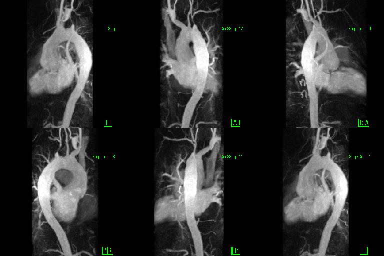
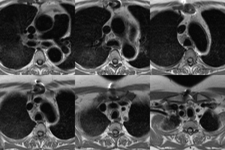
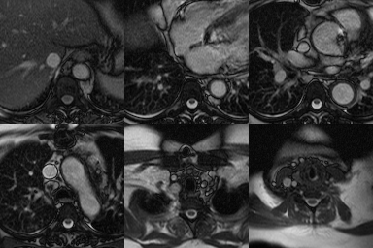
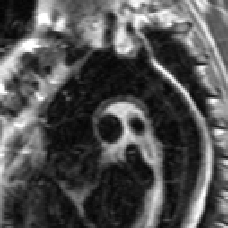
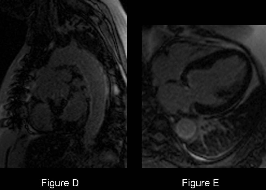
Selected images chest MRA / MRI. 3-D MRA (Fig. A) reveals luminal narrowing of the brachiocephalic and left common carotid artery and near complete occlusion of the left subclavian artery which fills via the left vertebral artery. Note the small left common carotid artery aneurysm. Axial cine HASTE (black-blood) (Fig. B) chest MRI images demonstrate marked narrowing of the left subclavian artery by a thick collar of intermediate signal intensity. Transverse, ascending and proximal descending and distal thoracic aorta likewise show a thickened wall of intermediate signal intensity contrasted with the low signal aortic lumen. Similar findings are seen on the axial SSFP MRI images (Fig. C). Note the signal drop-out from the median sternotomy wires and aortic valve replacement. Incidental note is made of partial anomalous venous drainage of the right upper lobe into the superior vena cava. 10-minute delayed post-contrast sagittal oblique (Fig. D) and 4-chamber (Fig. E) images show peripheral enhancement of the thickened aortic wall. Left subclavian steal was confirmed on cine VENC sequences (not illustrated) Diagnosis: Takayasu Arteritis
Differential Diagnosis
- Large-vessel vasculitides of other etiologies
Discussion
Background
Takayasu arteritis (aka pulseless disease; aortitis syndrome; idiopathic medial aortopathy) is an idiopathic granulomatous inflammation of the large arteries that may affect the aorta, its great vessels, and the pulmonary arteries. Marked intimal proliferation and fibrosis of the media and adventitia eventually lead to stenosis, occlusion, and, occasionally, post-stenotic dilatations and aneurysm formation. The inflammatory process tends to be segmental with a patchy distribution. Four major types have been described (Table-1).
Table-1. Takayasu Arteritis: Classification based on Vessel Involvement
Type Vessel Involvement Type I (Classic Pulseless) Brachiocephalic Artery + Carotid Arteries + Subclavian Arteries Type II Combination of Type I + III Type III (Atypical Coarctation) Thoracic and Abdominal Aorta distal to Arch & Great Vessels Type IV (Dilated) Extensive Dilatation of entire Aorta & Great Vessels The most common type is type III (65% of patients). The most commonly involved vessels include: left subclavian artery (50%); left common carotid artery (20%); brachiocephalic trunk; renal arteries; celiac trunk; superior mesenteric artery; and pulmonary arteries (50%). Infrequently, the axillary, vertebral, coronary, and iliac arteries are involved.
Etiology
Nonspecific, cell-mediated inflammatory process of unknown etiology.
Clinical Findings Takayasu arteritis most commonly occurs in women; the female-to-male ratio is 8:1. 90% of patients are younger than 30-40 years. C linical manifestations can be divided into 2 phases: early (prepulseless) phase - systemic symptoms (e.g., low-grade fever; malaise; weight loss; fatigue; tachycardia, pain adjacent to inflamed arteries [carotodynia]); and a late (pulseless or occlusive) phase- the most common symptom being related to vascular stenosis with diminished or absent pulses (96% of patients), often associated with limb claudication and blood pressure discrepancies. Not uncommonly, the disease is recurrent, leading to the coexistence of these phases simultaneously.
Imaging Findings
Chest Radiography
- Often normal
- Widened superior mediastinum
- Focal oligemia lung parenchyma
- Premature aortic calcification in younger patient population
MDCT / MRI
MRI modality of choice: avoids ionizing radiation exposure in young women
Early Stage
- Vessel wall thickening; crescentric; circumferential; irregular
Late Stage
- Vessel wall thickening; crescentric; circumferential; irregular (Fig. B-E)
- Luminal changes (Fig. A-E)
- Luminal narrowing, aneurysmal dilatation, and occlusion (Fig. A-E)
- Subclavian steel phenomenon may develop (Fig. A)
- Contrast enhancement of thickened vessel walls: some degree of active disease is present (Fig. D; E)
- MRI allows assessment of pressure differentials across stenotic lesions (VENC sequences)
- Mural calcium deposition in vessel walls in chronic phases
PET-FDG
- Useful indicator of vessel wall inflammation
- Intensity of FDG accumulation decreases in response to therapy
Treatment
- Early corticosteroid therapy: may lead to clinical improvements; subside active inflammatory phase; control or slow progression of disease
- Methotrexate and intravenous cyclophosphamide: glucocorticoid-resistant arteritis
- Angioplasty: generally contraindicated during acute phase of disease; may be successful once acute phase has abated
Prognosis
- Mortality; usually from vascular complications (e.g., hypertension, stroke, and aortic insufficiency
CAVEATS
- Takayasu arteritis affects almost exclusively patients younger than 40 years, involves primarily the aorta and its great vessels, and generally spares the cranial arteries
- Takayasu arteritis is the only form of aortitis that produces both stenosis and occlusion of the aorta
- Unilateral pulmonary artery occlusion can occur in advanced cases; Takayasu arteritis should be considered in cases of chronic pulmonary artery obstruction of unknown origin
Selected Readings
- Castaner E, Alguersuari A, Gallardo X, Andreu M, Pallardó Y, Mata JM, Ram?rez J. When to Suspect Pulmonary Vasculitis: Radiologic and Clinical Clues. RadioGraphics 2010; 30: 33-53.
- Engelke C, Schaefer-Prokop C, Schrig E, Freihorst J, Grubnic S, Prokop M. High-Resolution CT and CT Angiography of Peripheral Pulmonary Vascular Disorders. RadioGraphics 2002; 22: 739-764.
Original case written by its authors at Virginia Commonwealth University and published at this address as part of a weekly teaching collection. Reproduced here as an archive.