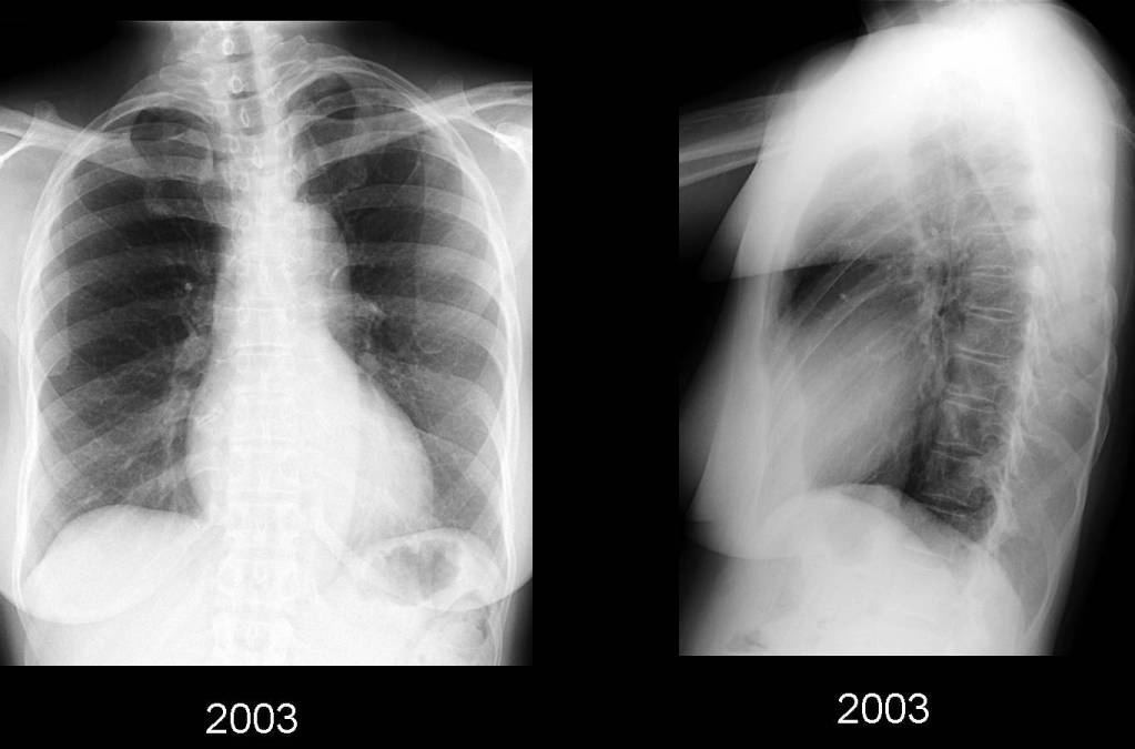
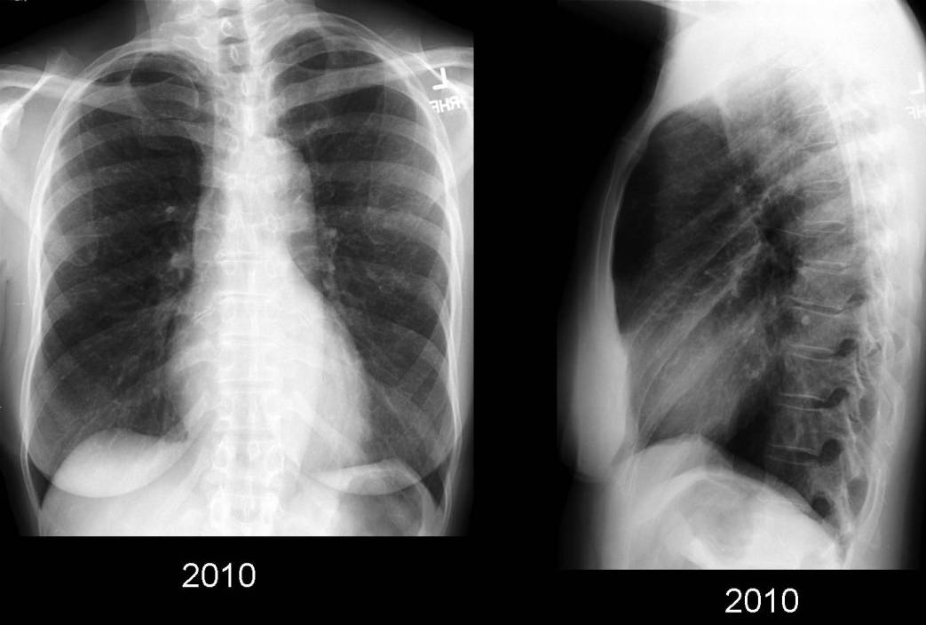
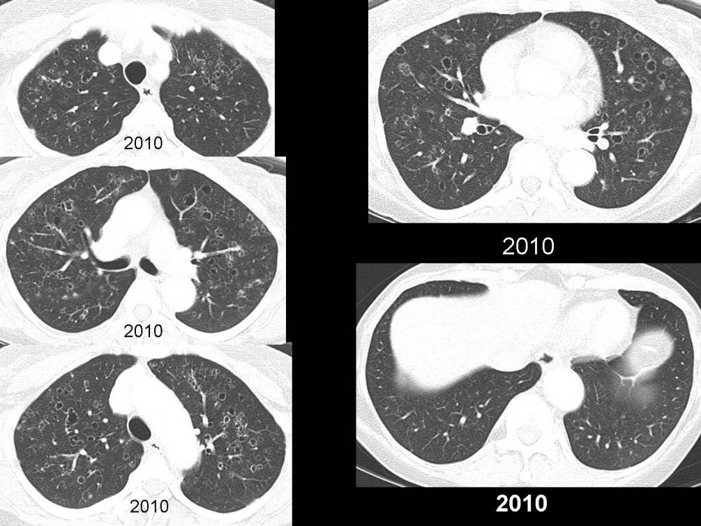
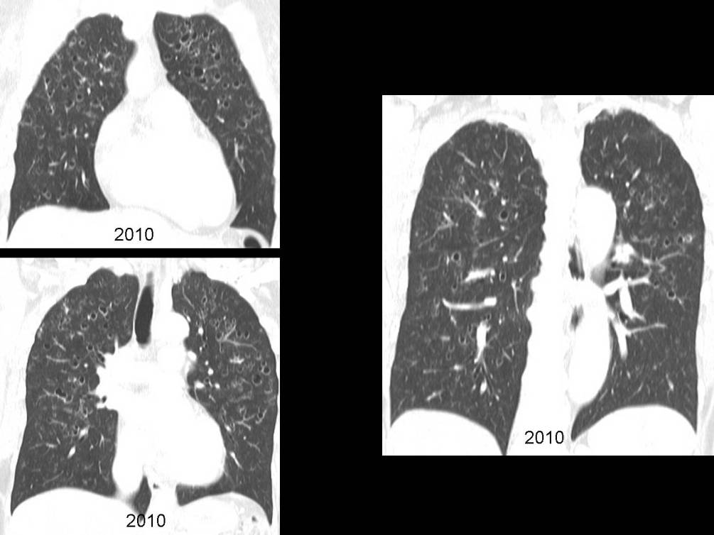
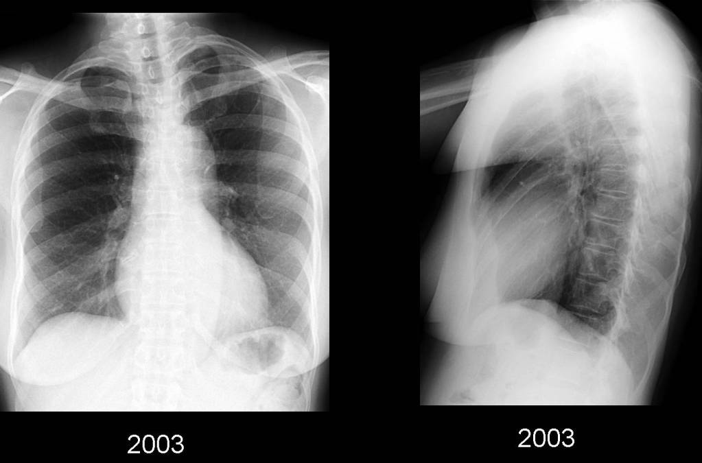
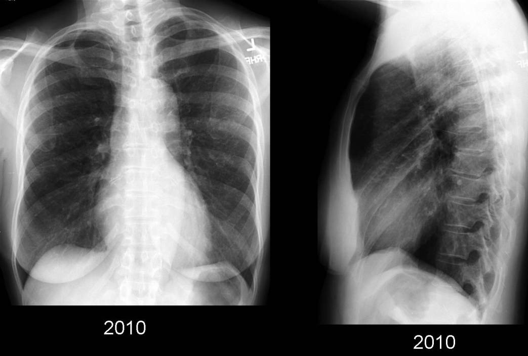
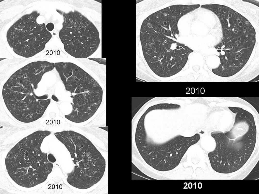
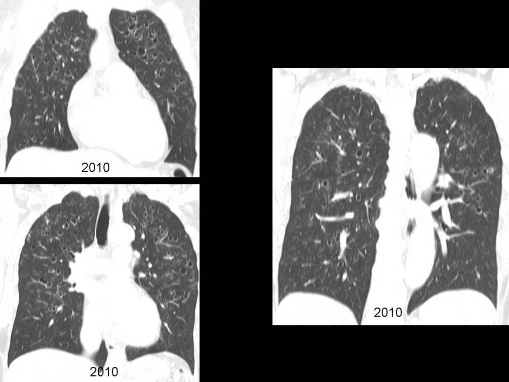
PA and lateral chest radiographs (2003) show preserved-to-slightly hyper-expanded lungs with a background of subtle reticular opacities most pronounced in the mid-and upper lung zones. Subsequent PA and lateral chest radiographs (2010), seven years later, show progression of this disease process and development of variable-sized lucent lesions in the same distribution. No lymphadenopathy, pleural effusion, or pneumothorax is seen. Axial and coronal reformatted Chest CT images performed at the same time (2010) confirm the presence of predominantly upper and mid- lung variable-sized centrilobular and peribronchial nodular opacities and cystic lesions. The lesions spare the posterior-basal segments of the lower lobes. Many of the cystic lesions demonstrate varying wall thickness and some cysts appear to coalesce and have bizarre configurations. The cystic lesions are parenchymal-based and clearly separate from the bronchi and bronchioles. Diagnosis: Pulmonary Langerhans’ Cell Histiocytosis (PLCH)
Clinical Pearl: Anatomic distribution of the disease with sparing of the posterior-basal segments of the lower lobes and a positive long-standing history of significant tobacco abuse.
Differential Diagnosis
- Lymphangioleiomyomatosis (LAM)
- Females in reproductive years
- Spherical cysts uniformly distributed throughout lungs
- Nodules uncommon
- Chylous effusions
- Laryngotracheal Papillomatosis
- Laryngeal and tracheal nodules
- Cysts usually in dorsal aspect of lower lobes
- Lymphocytic Interstitial Pneumonia (LIP)
- Most commonly seen in Sjögren syndrome
- No predilection for upper lobes
- Emphysema
- Pneumocystis Jiroveci Pneumonia
- Immunocompromised-immunosuppressed patients
- Cysts occur in areas of ground-glass attenuation
- Mycobacterium tuberculosis
- Cavities may have air-fluid levels; uncommon with PLCH
- (+) sputum for acid-fast bacilli (AFB)
- Birt-Hogge-Dubé syndrome
- Cutaneous manifestations (fibrofolliculomas, trichodiscomas/angiofibromas, perifollicular fibromas, and acrochordons)
- Pulmonary cysts; history of pneumothorax,
- Various types of renal tumors; most commonly oncocytoma
- Sarcoidosis
- Hypersensitivity Pneumonitis
- May demonstrate upper lobe predominance with sparing of costophrenic angles
- Nodules identical to PLCH
- Cysts usually smaller in number than PLCH
- Septic emboli
Discussion
Background
Pulmonary Langerhans’ Cell Histiocytosis (formerly known as pulmonary eosinophilic granuloma; pulmonary histiocytosis X) is a diffuse, chronic, progressive interstitial disease of lungs, strongly associated with cigarette smoking (90% of patients). PLCH is possibly the result of an allergic reaction to a constituent of cigarette smoke. The disease is characterized by multiple granulomata throughout the lungs that contain Langerhans’ cells. In addition to lung involvement, this disease can involve multiple other organ systems including bone (lytic lesions), pituitary gland (diabetes insipidus), mucous membranes, skin, lymph nodes, and liver.
Clinical Findings:
PLCH is primarily found in young to middle aged smokers, with an equal prevalence among males and females. Presenting symptoms include nonproductive cough, dyspnea, chest pain, fatigue, fever, and weight loss. 25% of patients may be asymptomatic at the time of diagnosis, with incidental discovery of the disease. Pneumothoraces occur in 25% of patients throughout the course of their disease. Bone lesions occur in approximately 10% of patients, with ribs being the most common site.
Imaging Findings
Chest Radiography
- May be normal
- Upper and mid-lung zone reticulonodular opacities with relative sparing of the lung bases
- Preserved or increased lung volumes
- Spontaneous pneumothorax; may be recurrent
- Bone involvement, especially rib lesions may be detected
CT
- More sensitive than conventional radiography
- Combination of nodules and cysts most common
- Lesions primarily located in upper and mid- lung zones; spare posterior-basal segments of lower lobes
Nodules
- Indistinct; irregular or stellate; smooth bordered; 1-10 mm; may be > 1.0 cm
- Profusion of nodules varies from isolated lesion to innumerable
- Progression of nodules to cavitary nodules to thick- and thin-walled cysts
- Centrilobular or peribronchial distribution
Cysts
- 1-3 cm in diameter
- Thin- or thick- walls
- May be the sole manifestation
- Spherical; lobulated; septated; partially septated; confluent; or bizarre in morphology
- End-stage disease: cysts may be so severe confused with end-stage emphysema
Treatment
- Primary treatment: cessation of smoking
- Progressive disease despite smoking cessation: steroids
- Advanced or refractory disease: lung transplantation; PLCH can recur in transplanted lung
Prognosis:
- Variable: complete remission to respiratory failure
- Mortality rate: 5%; worse in men; elderly; patients with recurrent pneumothoraces
- May resolve, stabilize, or progress to an end-stage fibrotic appearance
- ¾ patients will resolve or stabilize
Suggested Readings
- Abbott GF, et al. Pulmonary Langerhans’ cell histiocytosis. RadioGraphics 2004; 24:821-841.
- Brauner MW, et al. Pulmonary Langerhans’ cell histiocytosis: evolution of lesions on CT scans. Radiology 1997; 204; 497-502.
- Gurney JW, Winer-Muram HT, Rosado-de-Christenson ML, Mohammed TLH, Abbott GF, Maier MH. Pulmonary Langerhans Cell Histiocytosis. In: Specialty Imaging: HRCT of the Lung. Amirsys, Salt Lake City, Utah; 2009: 264-269.
- Parker MS, et al. Case 133: Pulmonary Langerhans’ Cell Histiocytosis in Teaching Atlas of Chest Imaging . Thieme, New York. 2006. 507-509.
Original case written by its authors at Virginia Commonwealth University and published at this address as part of a weekly teaching collection. Reproduced here as an archive.