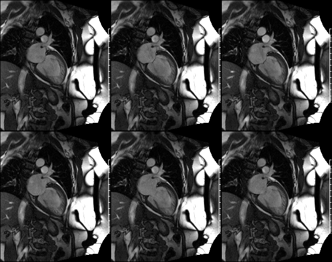
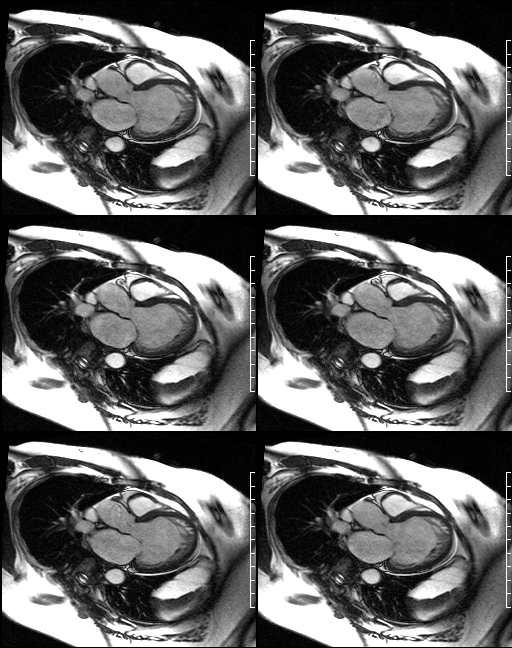
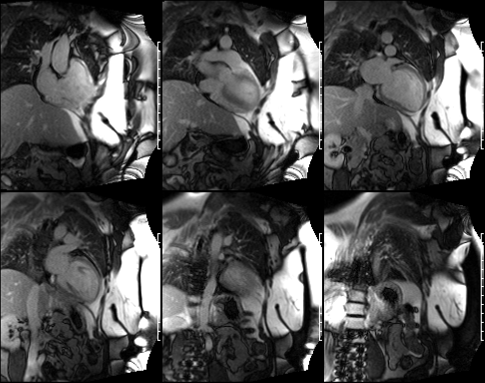
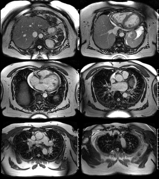
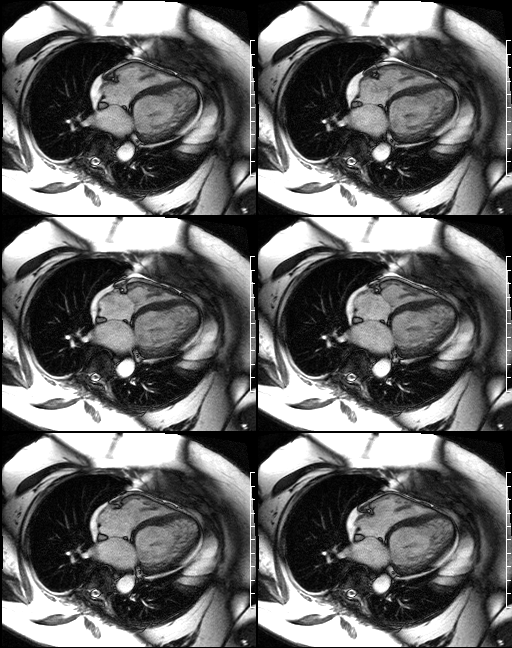
Selected Cardiac MRI. Cine GRE 2-chamber (Fig. A); Cine GRE 3-chamber (Fig. B); Cine Stacked GRE 2-chamber (Fig. C); Cine axial FISP (Fig. D); and Cine GRE-4 chamber (Fig. E) images demonstrate a markedly dilated left ventricular cavity estimated at 7.2 cm (end diastole) and 6.4 cm (end systole) with preserved wall thickness, estimated at 10 mm. There is diffuse global hypokinesis, worse at the apex, where extensive trabeculation is noted. The trabeculation has a "delaminated" appearance with only a thin residual compacted segment and extensive non-compacted segment. The ratio of non-compacted to compacted tissue is > 2.2:1. The ejection fraction was calculated at 11%. There is also mild right ventricular dilatation; mild mitral regurgitation, mild tricuspid regurgitation; and trace aortic insufficiency. Diagnosis: Diagnosis: Non-compaction of the myocardium
Differential Diagnosis None
Discussion
Background
Non-compaction or spongiform cardiomyopathy is a distinct cardiomyopathy characterized by excessive thickening of the myocardial wall which presents as a two-layered "delaminated" structure: a thin compacted epicardial layer and a substantially thicker non-compacted endocardial layer. The endocardial layer consists of prominent trabeculation and deep intratrabecular spaces which are in continuity with the left ventricular cavity. The radiologist should quantify the extent of non-compaction at the site of maximal wall thickness. In non-compaction, the ratio of non-compacted to compacted tissue is >2:1 during systole. Non-compaction is often associated with other congenital cardiac defects, but it is also seen in the absence of other cardiac anomalies.
Etiology
Myocardial non-compaction results from failure of normal myocardial trabeculated tissue to compact or transform from spongy to solid tissue during embryogenesis. Familial occurrence has been reported although the pattern of inheritance is not known. The American Heart Association's 2006 classification of cardiomyopathies considers non-compaction as a genetic cardiomyopathy. Mutations in LD B3 have been described in patients with this cardiomyopathy.
Clinical Findings
The clinical course is variable but may be complicated by progression to dilated cardiomyopathy and heart failure. Adults with non-compaction are more likely to suffer from heart failure and affected children more often experience symptoms related to depression of systolic function. Tachyarrhythmia which can lead to sudden death and intracardiac stasis and thrombus formation are also more common with non-compaction.
Imaging Findings
Cardiac MRI (Figure A-E)
- Dilated left ventricular cavity with preserved wall thickness
- Diffuse global hypokinesis, worse at the apex, where extensive trabeculation is noted
- Decreased ejection fraction
- Trabeculation has a "delaminated" appearance
- Ratio of non-compacted to compacted tissue is > 2:1 during systole
- +/- Right ventricular dilatation
- +/- Coronary microcirculatory disturbances with abnormal perfusion even in remote myocardial segments not involved with the disorder
Management
Medical Therapy
Similar to that of other types of cardiomyopathies
- ACE Inhibitors
- Beta Blockers
- Aspirin and/or other forms of anticoagulation
Surgical Therapy
- Pacemaker for patients at high risk of arrhythmia
- Transplant; severe cases of medical refractory heart failure
Prognosis
- Relatively new disease; potential impact on life expectancy is not fully understood at this time
- Long term prognosis is currently unknown
Caveats
- Arrhythmias are common in patients with ventricular non-compaction
- Atrial fibrillation has been reported in over 25% of adults with non-compaction
- Ventricular tachyarrhythmia's have been reported in as many as 47% affected patients
Selected Readings
- Grizzard JD, Judd RM, Kim RJ. Teaching File Case 46. In: Cardiovascular MRI in Practice: A Teaching File Approach. Springer-Verlag, London; 2008:146-147.
- Maron, Barry.; Towbin, Jeffrey.; Thiene, Gaetano; Antzelevitch, Charles; Corrado, Domenico.; Arnett, D; Moss, AJ; Seidman, CE et al. (2006). Contemporary Definitions and Classification of the Cardiomyopathies (webpage). American Heart Association Journals (American Heart Association) 113 (14): 1807. doi : 10.1161/CIRCULATIONAHA.106.174287 . Accessed 14 April 2010.
- Oechslin, Erwin; Jenni, Rolf (2005) (webpage). Non-compaction of the left ventricular myocardium-From Clinical Observation to the Discovery of a New Disease. http://www.touchcardiology.com/compaction-leftventrivcular-a352-3.html.%20Accessed%2014%20April%202010 .
Original case written by its authors at Virginia Commonwealth University and published at this address as part of a weekly teaching collection. Reproduced here as an archive.