Patent Ductus Arteriosus (PDA); Eisenmenger Physiology with Pulmonary Artery Hypertension
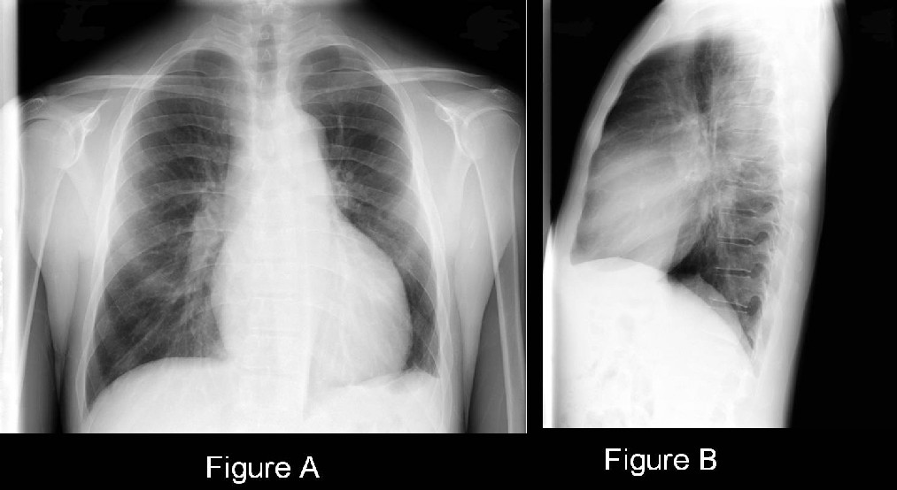
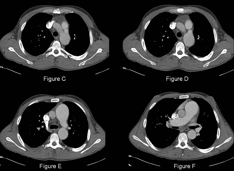
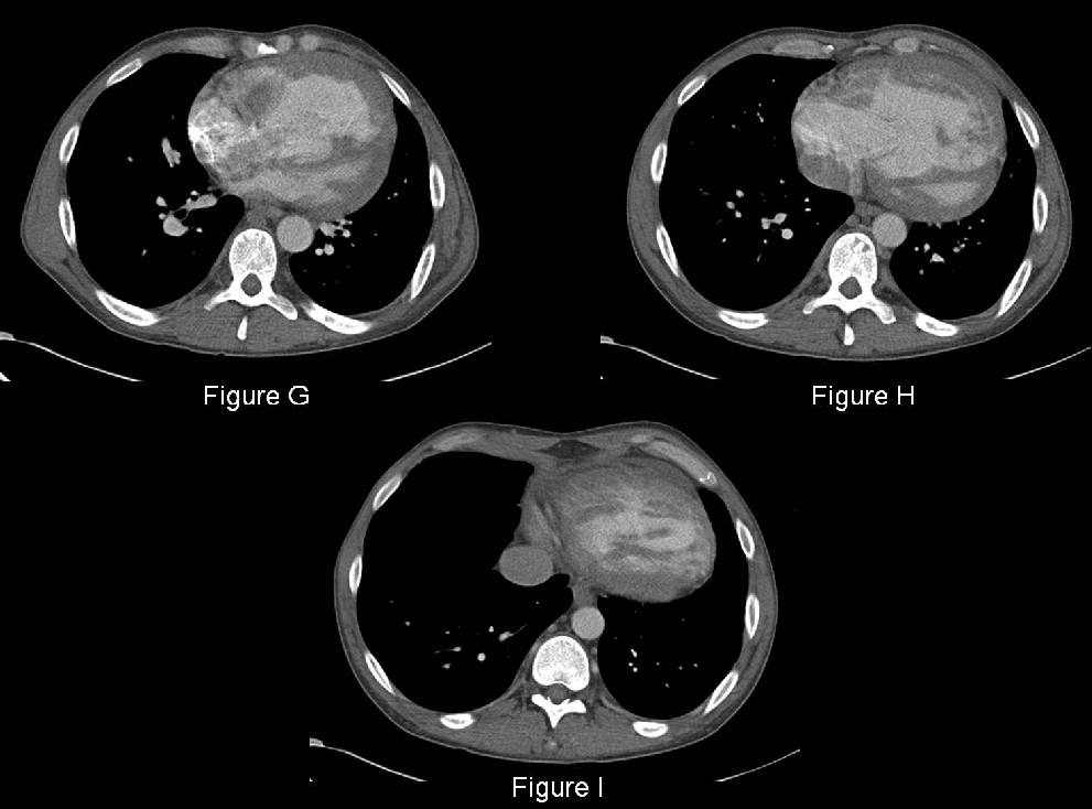
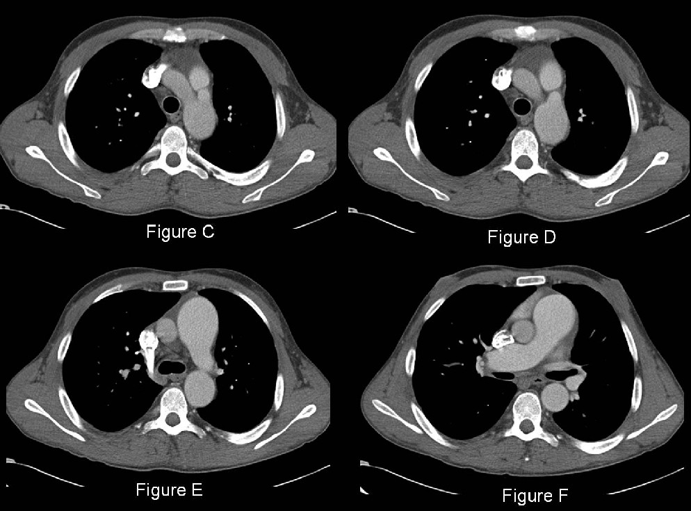
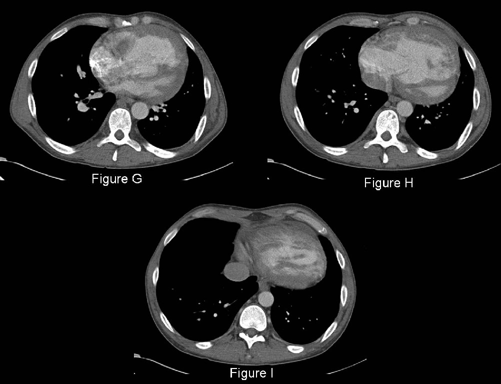
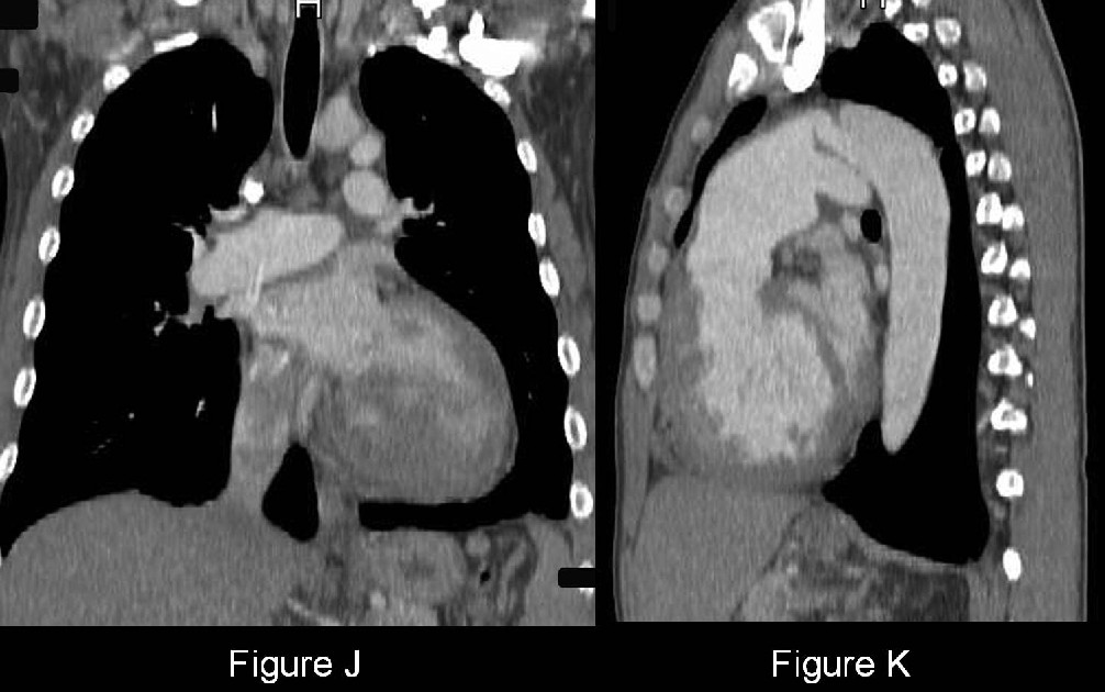
PA (Fig. A) and lateral (Fig. B) chest radiographs demonstrate an enlarged cardiomediastinal silhouette and central pulmonary arteries with pruning of the peripheral vessels. The lateral exam (Fig. B) demonstrates encroachment of the retrosternal clear space by an enlarged right ventricle. Contrast-enhanced axial CT images (Fig. C-I) confirm the presence of an enlarged heart, right ventricular dilatation and right ventricular hypertrophy in particular (Fig. G-I). Note the thickened right ventricular wall. Compare the relative size of the right ventricle to that of the smaller and posteriorly displaced left ventricle. The interventricular septum is displaced toward the left ventricular cavity from the increased right heart pressures. The main pulmonary artery is dilated relative to the ascending aorta (Fig. D-F). An enhancing tubular structure is seen between the proximal descending aorta and main pulmonary artery (Fig. C; D; J; K). The descending thoracic aorta is enlarged relative to the ascending aorta and there is asymmetric enhancement of the ascending aorta compared to the descending aorta proximal to this tubular structure (Fig. C-F) suggesting disproportionately elevated pulmonary artery pressures relative to systemic pressures. Diagnosis: Patent Ductus Arteriosus (PDA); Eisenmenger Physiology with Pulmonary Artery Hypertension
Differential Diagnosis
None
Discussion
Background
The ductus arteriosus is a pathway of fetal circulation connecting the pulmonary artery and aorta. It functions as a bypass tract for intrauterine blood flow away from the lungs. Normally, the ductus arteriosus functionally closes by 48 hours following birth and anatomically by 4 weeks of life. When it fails to close, it is referred to as a patent ductus arteriosus (PDA).
Etiology
The incidence of failure of ductus arteriosus closure is highest among premature infants. It is more common in females than in males, is associated with surfactant deficiency, and can be seen with maternal rubella. With increased survival of premature infants, there has been an increase in the amount of patients with all types of congenital heart disease surviving into adulthood.
Clinical Findings
Clinical symptoms typically do not present at birth because the transition from fetal circulation is not immediate. Dyspnea and cyanosis present more than a week after birth. PDA’s are generally small in asymptomatic adults and as such, found incidentally. Pursuit of this diagnosis is uncommon until the patient becomes symptomatic. Both shunt size and the difference between systemic and pulmonary vascular resistance determine the physiologic consequence of PDA in adults. This is because these factors determine the degree of left-to-right shunting. With a small PDA, the degree of shunting may be trivial and the patient remains asymptomatic. Larger PDA’s cause a continuous murmur best appreciated over the upper left parasternal border. There may also be a widened pulse pressure. ECG in the setting of a large shunt may reveal signs of left atrial dilatation and left ventricular strain. This patient exhibited differential cyanosis (i.e., upper extremities appeared pink and well perfused, while lower extremities appeared cyanotic) reflecting the onset of Eisenmenger physiology (i.e., shunting of blood flow back to the lung causing pulmonary artery hypertension and shunt reversal). The pulmonary arterial pressure (PAP) was reported measured in excess of 130 mmHg (normal 12-16mmHg). In such cases, the upper body continues to receive well oxygenated blood via the aortic branch vessels. However, the ensuing right-to-left shunt sends deoxygenated blood from the pulmonary artery, through the patent ductus (located beyond the branch vessels) to the descending aorta.
Imaging Findings
Conventional Radiography
Infants and Young Children
- Enlarged hilar and segmental vessels (when not obscured by the thymus)
- Perihilar pulmonary edema (when shunt > 2:1)
- May progress to generalized alveolar edema
- Cardiothoracic ratios > 0.55
- +/- left atrial enlargement on lateral exams
Adults
- May appear normal depending on shunt size
- Enlarged pulmonary artery segment (Fig. A and B)
- Enlarged aorta (Fig. A and B)
- Enlarged right ventricle (Fig. B)
- +/- left atrial enlargement
- Increase in pulmonary blood flow (less flow directed towards LUL)
- “Ductus bump” sign – localized dilatation adjacent to descending aorta next to the arch at the entrance of the ductus; may silhouette aortic pulmonary window
- Calcified ductus arteriosus calcifications (“railroad track” sign)
- Pruning of peripheral vasculature with development of shunt reversal and Eisenmenger physiology (Fig. A and B)
MDCT/MRI
- Confirms the conventional radiography findings (Fig. C-K)
- Right ventricular dilatation-hypertrophy (Fig. G-K)
- +/- left ventricular enlargement
- +/- left atrial enlargement
- Dilatation of the main pulmonary artery (Fig. C-F)
- Patent vascular channel from the distal aortic arch to the left side of the main pulmonary artery (Fig. C; D; J; K)
- +/- ductus calcification
- ECG-gated MDCT can be used to accurately describe the surface anatomy and internal structure, as well as provide 3D reconstructions.
- Phase Encoding Pulse Sequences: assess and quantify the degree of shunting and gradient across the PDA.
Treatment
In children, indomethacin is administered. This inhibits prostaglandin E1 which is responsible for keeping the ductus open. Catheter closure with thrombogenic coils or placement of an umbrella device is the procedure of choice for small PDA’s. For large PDA’s where catheter closure is not possible, surgical ligation is preferred.
Prognosis
Without treatment, the overall mortality for PDA in adults regardless of size is 1.8% per year. Patients with a small PDA’s usually remain asymptomatic. Patients with a large PDA may develop heart failure and pulmonary hypertension. Regardless of size, complications such as endarteritis, endocarditis, and embolic events may occur. It is thus generally recommended that patients discovered to have PDA undergo closure procedures, even if the PDA is small.
References
- Akutsu Y, Gokan T, Seino N, Kaneko K, Kodama Y, Kobayashi Y. Multidetector computed tomography in an adult patient with silent patent ductus arteriosus. Clin Cardiol. 2009 Dec; 32(12):E85.
- Cassidy HD, Cassidy LA, Blackshear JL. Incidental discovery of a patent ductus arteriosus in adults. J Am Board Fam Med. 2009 Mar-Apr; 22(2):214-8.
- Goitein O, Fuhrman CR, Lacomis JM. Incidental finding on MDCT of patent ductus arteriosus: use of CT and MRI to assess clinical importance. AJR Am J Roentgenol. 2005 Jun; 184(6):1924-31.
- Morgan-Hughes GJ, Marshall AJ, Roobottom C. Morphologic assessment of patent ductus arteriosus in adults using retrospectively ECG-gated multidetector CT. AJR Am J Roentgenol. 2003 Sep; 181(3):749-54.
- Supriya Sharma, Atul C. Mehta and Peter B. O'Donovan. Computed Tomography and Magnetic Resonance Findings in Long-standing Patent Ductus; Case Reports Angiology 1996; 47; 393.
- Wiyono SA, Witsenburg M, de Jaegere PP, Roos-Hesselink JW. Patent ductus arteriosus in adults: Case report and review illustrating the spectrum of the disease. Neth Heart J. 2008 Aug; 16(7-8):255-9.
Original case written by its authors at Virginia Commonwealth University and published at this address as part of a weekly teaching collection. Reproduced here as an archive.