Differential Diagnosis
Unilateral Opaque Thorax
- Pleural effusion
- Fibrothorax
- Primary and secondary pleural malignancy
- Pneumonia
- Atelectasis
- Tuberculosis
- Pulmonary agenesis or hypoplasia
- Pneumonectomy
- Diaphragmatic hernia
- Chest wall deformity
- Thoracic spine scoliosis
The differential diagnosis can be further narrowed by noting the relative position of the trachea air column and mediastinum relative to the opaque thorax as follows:
Unilateral Opaque Thorax with Midline Trachea
- Extensive parenchymal consolidation
- Malignant pleural disease (e.g., malignant mesothelioma)
Unilateral Opaque Thorax with Contralateral Tracheal Displacement
- Massive hydrothorax of various etiologies
- Diaphragmatic hernia
- Other pleural space occupying masses
Unilateral Opaque Thorax with Ipsilateral Tracheal Displacement
- Central obstructing tumor with post-obstructive lung collapse
- Uncomplicated lung collapse (e.g., obstructing foreign body, mucus plug)
- Following pneumonectomy
- Pulmonary agenesis or hypoplasia
Discussion
Background
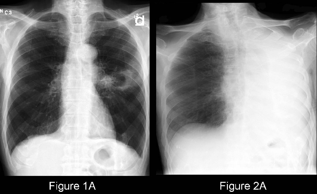
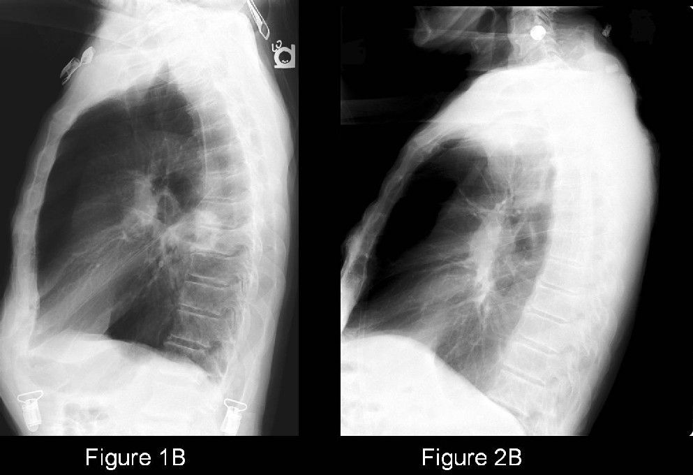
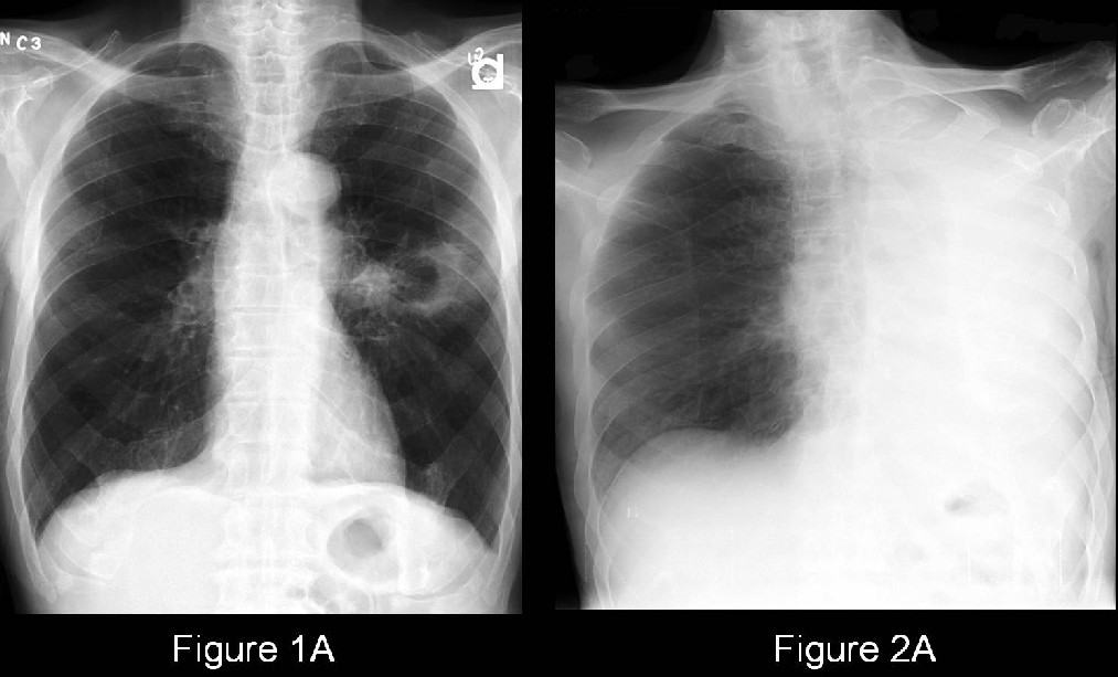
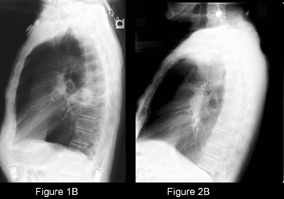
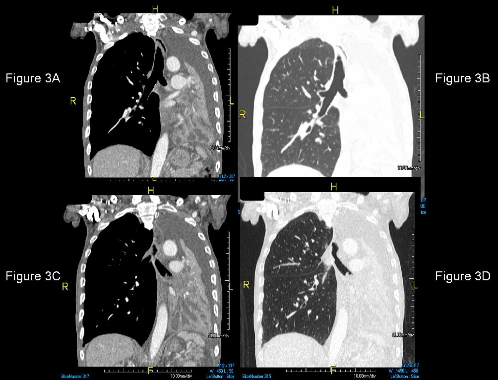
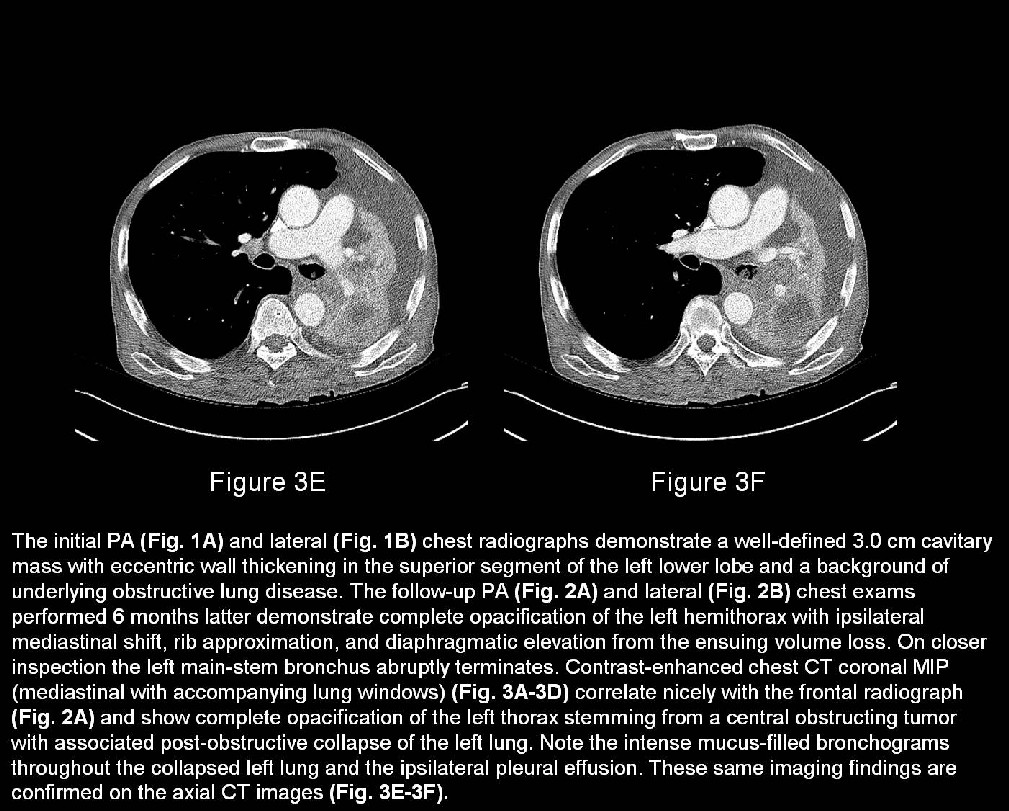
The baseline chest radiographs (Fig. 1A and 1B) revealed a well-defined cavitary mass with eccentric wall thickening. As a general rule, the more irregular or thickened the wall of a cavitary lesion becomes, the more likely the lesion is malignant. The most common cavitary neoplasm in the lung is squamous cell carcinoma. Squamous cell carcinoma is also the most common lung neoplasm to present as a central obstructing lesion, followed by small cell carcinoma. Over the 6 months between the 2 studies, the tumor progressed until there was complete obstruction of the main-stem bronchus and resultant post-obstructive collapse of the left lung (Fig. 2 and 3).
Treatment
- The tumor was non-resectable in this particular case
- Laser therapy partially restored some patency to the left upper lobe
- Palliative radiation and chemotherapy
Caveats:
- The more irregular or thickened the wall of a cavitary lesion becomes, the more likely the lesion is malignant.
- The broad differential diagnosis for a unilateral opaque thorax can be further narrowed by noting the relative position of the trachea air column and mediastinum relative to the opaque thorax.
- Squamous cell accounts for 25-40% of all lung cancers; usually develops in proximal airways (centrally); the most likely cell type to cavitate; and the cell type least likely to metastasize distantly
Selected Readings
Lange S, Walsh G. Radiology of Chest Diseases. Radiographic Signs and Differential Diagnosis. In:
Radiology of Chest Diseases. New York, NY: Thieme Scientific and Medical Publishers, 2007:301-304.
Original case written by its authors at Virginia Commonwealth University and published at this address as part of a weekly teaching collection. Reproduced here as an archive.