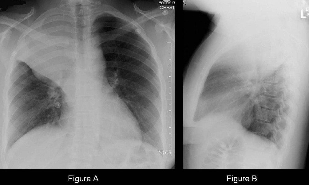
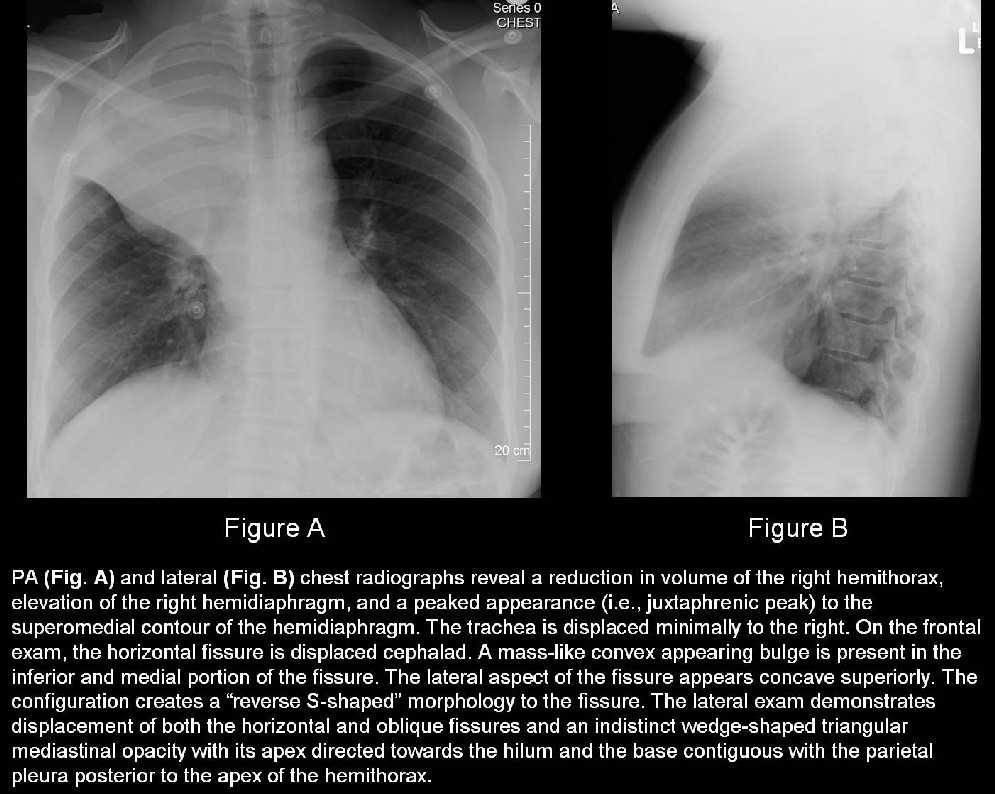
Complicated right upper lobe atelectasis caused by a large hilar mass forming the “reverse S sign of Golden; lymphoma
Differential Diagnosis
- Post-obstructive upper lobe atelectasis from an endobronchial mass (e.g., primary and secondary neoplasia)
- Post-obstructive upper lobe atelectasis from extrinsic bronchial compression by reactive or neoplastic lymphadenopathy
Discussion
Background
In non-complicated right upper lobe atelectasis (e.g., aspirated foreign body, mucus plug), the inferior border of the horizontal fissure should be concave on frontal and lateral radiography. Complicated right upper lobe atelectasis caused by a hilar mass (e.g., small cell carcinoma, squamous cell carcinoma, lymphoma, lymphadenopathy, etc) is often associated with a convex bulge in the medial aspect of the horizontal fissure. The lateral aspect of the horizontal fissure is appropriately concave. The result is a “reverse S-shaped” configuration to the horizontal fissure (i.e., reverse “S” sign of Golden), a sign highly suggestive of a central neoplasm as the etiology of the atelectasis.
Clinical Findings
Affected patients may present with cough, weight loss, hypoxemia, progressive dyspnea, and hemoptysis, diminished or absent breath sounds over the affected area, and various paraneoplastic syndromes depending upon the etiology of the obstructing lesion.
Imaging Findings
- Opaque right upper thorax (Fig. A and B).
- Direct and indirect signs of right hemithoracic volume loss (Fig. A and B).
- Convex bulge medial aspect of displaced horizontal fissure (Fig. A and B).
- Central hilar mass lesion or extrinsic bronchial compression by lymphadenopathy (Fig. A and B).
Management
- Prompt bronchoscopic evaluation and biopsy to determine etiology of the complicated lobar atelectasis and establish a tissue diagnosis.
- Surgical resection when possible.
- Endobronchial stent deployment in select cases.
- Palliative chemotherapy and / or radiation therapy in unresectable cases.
Prognosis
- Dependent on the stage at presentation in cases of neoplasia
Caveats
- The “reverse S sign of Golden” is highly suggestive of a central bronchogenic carcinoma as the cause of the atelectasis on both radiography and CT and its presence on chest radiography should always prompt further investigation with bronchoscopy and / or CT. This sign is NOT seen in cases of uncomplicated atelectasis (e.g., aspirated foreign body, mucus plug).
- Although initially described for right upper lobe atelectasis, the sign is applicable to atelectasis of any lobe.
Suggested Readings
- Golden R. The effect of bronchostenosis upon the roentgen-ray shadows in carcinoma of the bronchus. Am J Roentgenol Radiat Ther 1925; 13:21-30.
- Proto AV, Tocino I. Radiographic manifestations of lobar collapse. Semin Roentgenol 1980; 15:117-173.
- Reinig JW, Ross P. Computed tomography appearance of Golden's "S" sign. J Computed Tomogr 1984; 8(3): 219-23.
- Woodring JH, Reed JC. Types and mechanisms of pulmonary atelectasis. J Thorac Imaging 1996; 11:92-108.
Original case written by its authors at Virginia Commonwealth University and published at this address as part of a weekly teaching collection. Reproduced here as an archive.