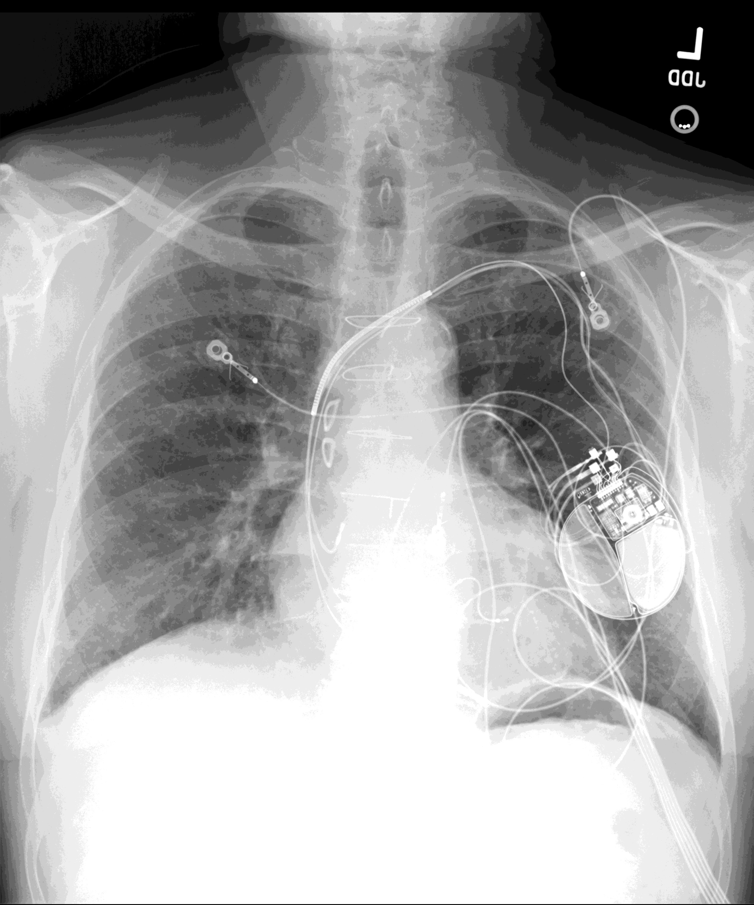
There are additional findings of median sternotomy, coronary artery bypass graft surgery, left coronary arterial stent, and an AICD. The nuclear medicine myocardial perfusion study demonstrates a fixed defect with the LV apex. The CT scan of the chest demonstrates a 4 x 3 cm calcified aneurysm of the apicoseptal left ventricle with adjacent thrombus formation. Answer Diagnosis: The lateral chest radiograph demonstrates a calcified aneurysm at the left ventricular cardiac apex
The lateral chest radiograph demonstrates a calcified aneurysm at the left ventricular cardiac apex
DIFFERENTIAL DIAGNOSIS
The differential diagnosis includes pericardial calcifications, calcified pericardial cyst, calcified left ventricular luminal thrombus, calcified LV pseudoaneurysm, and calcified cardiac neoplasm (teratoma).
DISCUSSION
Left ventricular aneurysm (LVA) is defined as a fibrotic region of the LV myocardium, with or without calcification, secondary to transmural infarction, which demonstrates dyskinetic motion. 95% of LVAs are due to myocardial ischemia, rarely trauma, Chagas disease, or sarcoidosis. LVA usually arises from the LV anterior wall/apex (85%) in the LAD territory. 15% of LVA arises from the inferior LV wall in the RCA territory. Lateral wall LVA, in the circumflex territory is rare. LVA may be the underlying cause of heart failure, arrhythmia, or arterial embolism.
Associated mural thrombus increases the risk of thromboembolism to 19% per year, and anticoagulation is required, as in this case. If no mural thrombus is present, then anticoagulation is typically not required.
REFERENCES
1. “Giant and Calcified Post-Infarction true Left Ventricular Aneurysm: What To Do?”, Kajita, Oliveira, Nicolau, Arq Bras Cardiology, 2016, March, 106(3), 259-262.
2. “Calcified Ventricular Aneurysm”, Oman Medical Journal, 2009, Oct: 24(4):303-305.
(Click for a larger image.)
Original case written by its authors at Virginia Commonwealth University and published at this address as part of a weekly teaching collection. Reproduced here as an archive.