Thoracic Imaging Archive
34 language editions

Archived case 15 · Sep 10-Sep 17, 2009

Intrathoracic Splenosis

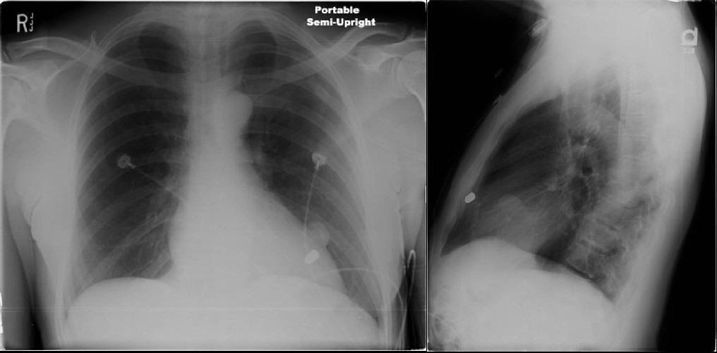
62-year-old-man with chest tightness, shortness of breath, and remote history of gunshot wound to the abdomen in the 1970’s.
Images

Radiograph 1 from archived case 15
Figure 1
Radiograph 2 from archived case 15
Figure 2
Radiograph 3 from archived case 15
Figure 3
Radiograph 4 from archived case 15
Figure 4
Radiograph 5 from archived case 15
Figure 5
Radiograph 6 from archived case 15
Figure 6

Diagnosis

Intrathoracic Splenosis

Differential Diagnosis

  • Pleural-based metastases
    • Various extrathoracic neoplasms
    • Lymphoma
    • Invasive thymoma
  • Mesothelioma
  • Pleural plaques
  • Lung metastases

Discussion

Background

This rare diagnosis represents the spontaneous autotransplantation of splenic tissue to various ectopic sites. It usually occurs as sequelae of penetrating or blunt traumatic injury to the spleen.

Etiology

Splenosis most frequently manifests with omental, peritoneal, and mesenteric implants in the abdominal cavity. However, implants of autotransplanted splenic tissue may also involve the retroperitoneum and liver surface. Intrathoracic splenosis may occur when fragments of disrupted splenic tissue gain access to the thorax either via a diaphragmatic tear (as in this particular case) or through diaphragmatic foramina, with subsequent implantation along the visceral or parietal pleura, interlobar fissure, or pericardium of the left hemithorax. These ectopic splenic implants then derive their own blood supply and grow histologically into mature, functional splenic tissue.

Clinical Findings

Intrathoracic splenosis has been identified in patients 6-42 years following the traumatic insult. Thus, the inciting event may have been long since forgotten by the patient and not recognized or considered relevant by the affected patient’s current day practitoner. Most patients are indeed asymptomatic with these nodular opacities discovered on chest radiography obtained for unrelated reasons. Because intrathoracic splenosis manifests as pleural-based lesions, patients are often misdiagnosed with metastatic disease and undergo invasive biopsy or thoracotomy. Uncommonly reported symptoms include pleuritic chest pain and recurrent hemoptysis.

Pathology

  • Sessile or pedunculated reddish-blue nodules
  • 7 mm-to-several centimeters in diameter
  • Number from a few-to-several hundred

Imaging

Radiography

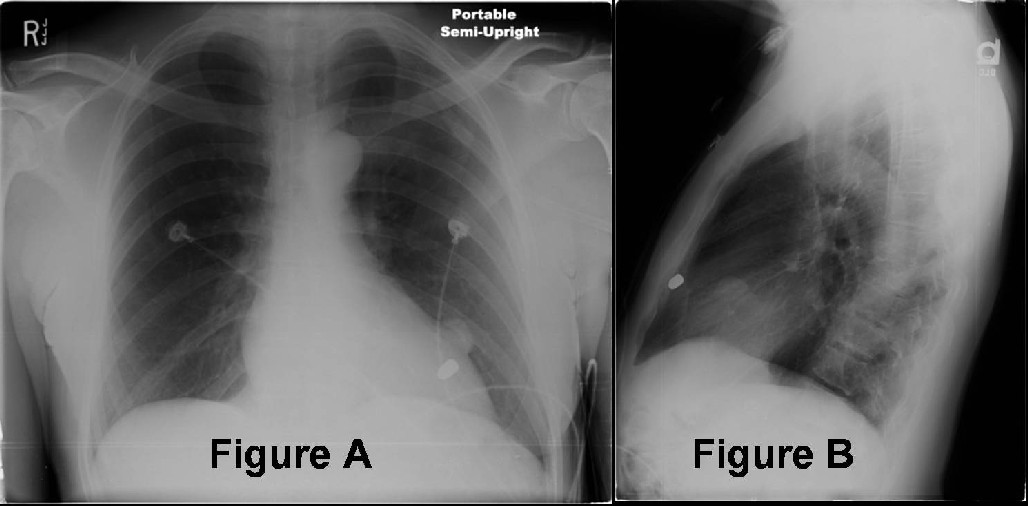
  • Single or multiple pleural-based non-calcified nodules (Fig. A and Fig. B)
  • Few millimeters to 7.5 cm in diameter (Fig. A and Fig. B)
  • Invariably left-sided (Fig. A and Fig. B)
  • Remote ribs fractures, irregularity of the ipsilateral diaphragm, retained shrapnel helpful clues to the diagnosis (Fig. A and Fig. B)

CT

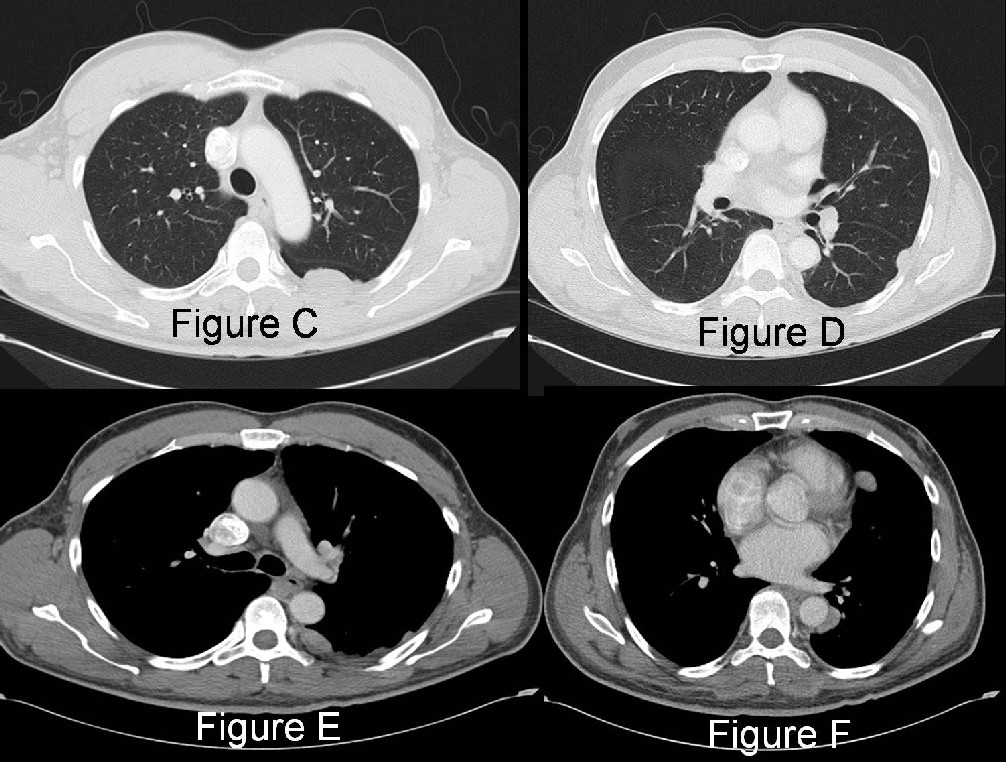
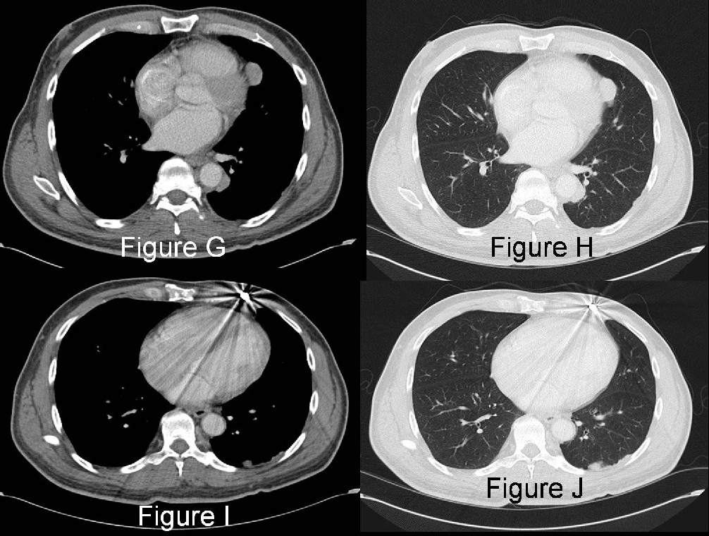
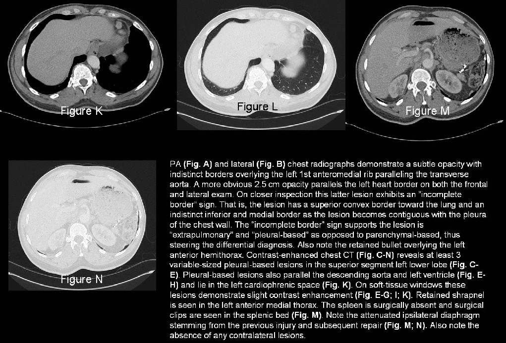
  • Single or multiple pleural-based non-calcified nodules (Fig. C-N)
    • Visceral or parietal pleura (Fig. C-N)
    • Paraspinal costovertebral pleura (Fig. E)
    • Interlobar fissures
    • Pericardial and/or mediastinal pleural reflections (Fig. E-H)
  • Few millimeters to 7.5 cm in diameter (Fig. C-N)
  • Invariably left-sided (Fig. C-N)
  • Measured attenuation similar to normal spleen (30-70HU) (Fig. E-G; I; K) or slightly lower

MRI

  • Comparable signal intensity to normal splenic tissue
  • T1WI: isointense to paraspinal muscles
  • T2WI: isointense to subcutaneous fat

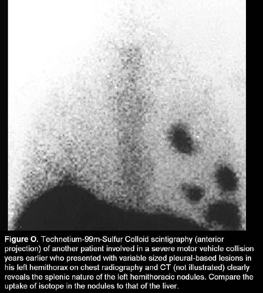
Nuclear Scintigraphy

  • Technetium-99m-tagged heated-red blood cells
  • 99m-Tc Sulfur Colloid
  • Indium-111-labeled platelets
  • Confidently establishes the radiologic diagnosis
  • Permits identification of the splenic nature of the nodules identified on chest radiography and CT as well as other potentially inconspicuous nodules (Fig. O)

Management

  • Surgical removal of the intrathoracic splenic tissue is not indicated in most cases and should be avoided

Prognosis

  • Removal of thoracic splenic tissue in a patient without functional abdominal splenic tissue may render the patient asplenic, increasing risk of infection.

CAVEATS

  • Although rare, the diagnosis of intrathoracic splenosis should be considered in a patient with left hemi-thoracic pleural-based nodules with a history of antecedent thoracoabdominal trauma and splenectomy or splenic injury.
  • Noninvasive diagnostic confirmation with nuclear scintigraphy may help patients avoid additional follow-up imaging studies, and/or invasive procedures including potential thoracotomy.

Suggested Readings

  1. Hagman TF, Winer-Muram HT, Meyer CA, Jennings SG. Intrathoracic splenosis: Superiority of Technetium Tc 99m Heat-Damaged RBC Imaging. Chest 2001; 120(6): 2097-2098.
  2. Moncada R, Williams V, Fareed J, et al. Thoracic splenosis. AJR Am J Roentgenol 1985; 144,705-706.
  3. Naylor MF, Karstedt N, Finck SJ, et al. Noninvasive methods of diagnosing thoracic splenosis. Ann Thorac Surg 1999; 68,243-244.
  4. White CS, Meyer CA. General case of the day. RadioGraphics 1998; 18, 255-257.

Filed under: Radiology, Medicine/Pulmonary

Original case written by its authors at Virginia Commonwealth University and published at this address as part of a weekly teaching collection. Reproduced here as an archive.

Back to the case index