Multiple myeloma with expansile rib lesions.
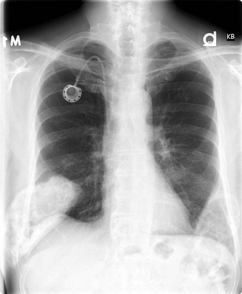
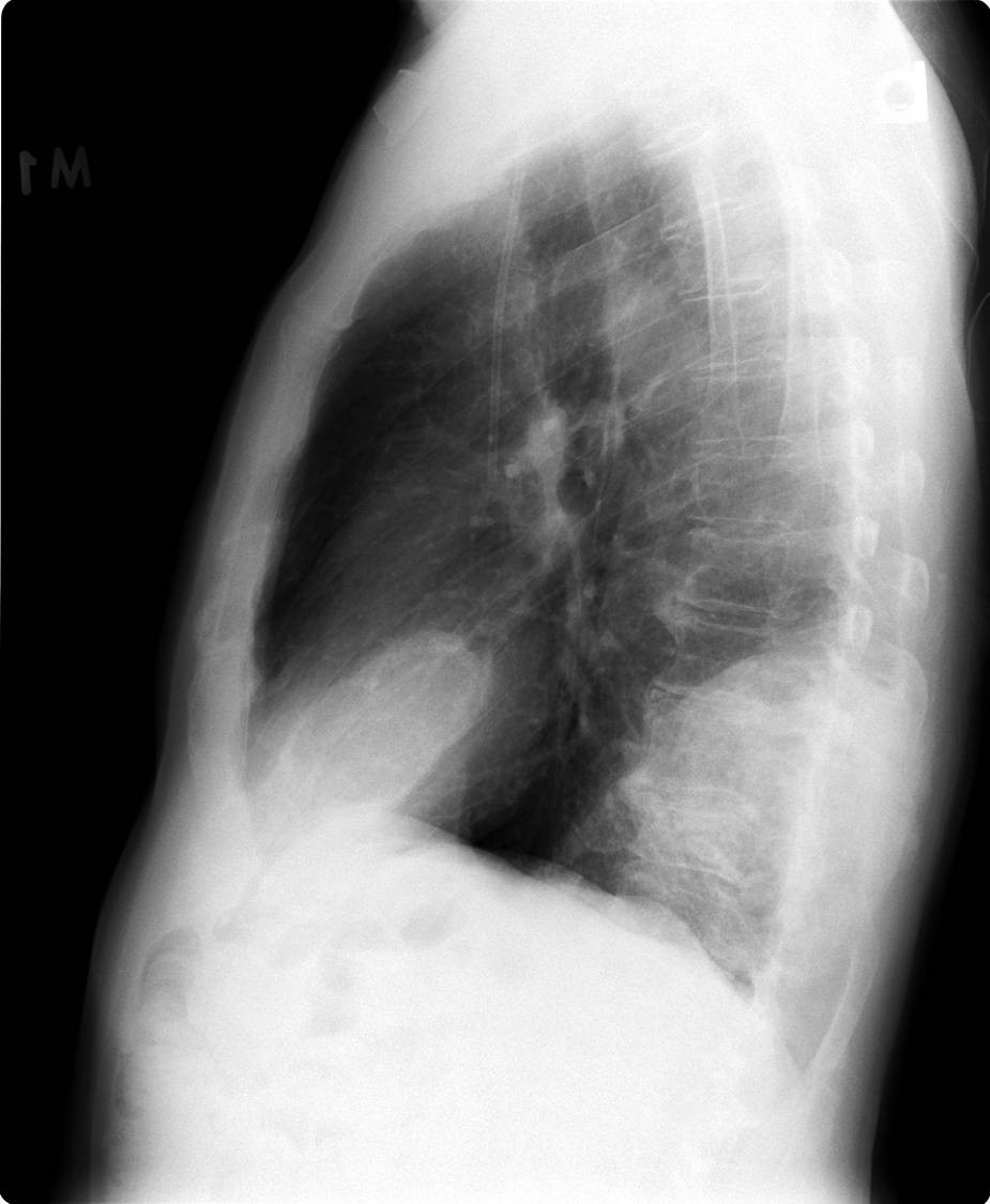
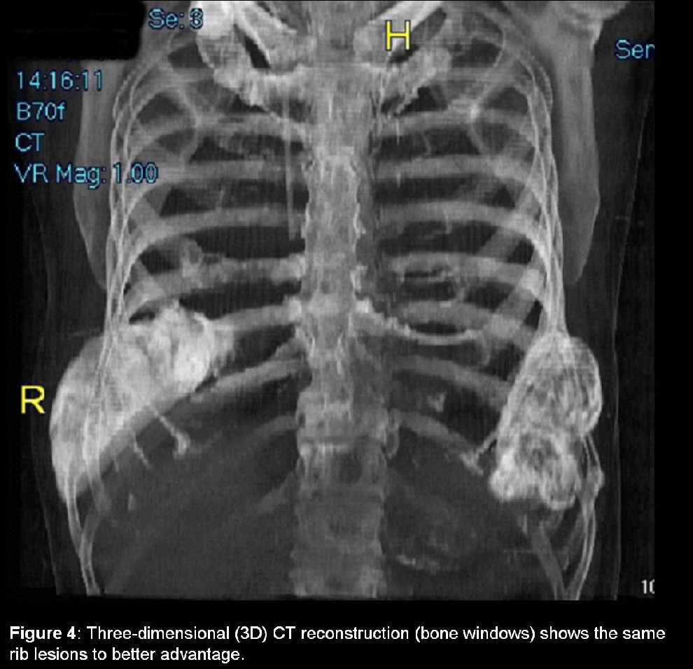
PA and lateral chest radiographs reveal two large, expansile, partially calcified masses arising from the right ninth posterolateral and left seventh anterolateral ribs, with destruction of the underlying bone. There are additional lytic lesions in multiple ribs, the right clavicle, and the thoracic spine. Diagnosis: Multiple myeloma with expansile rib lesions.
Differential Diagnosis:
- Benign bone tumors:
- Polyostic fibrous dysplasia
- Aneurysmal bone cyst
- Enchondroma
- Eosinophilic granuloma
- Chondroblastoma
- Chondromyxoid fibroma
- Giant cell tumor
- Hemangioma
- Extramedullary hematopoesis
- Malignant bone tumors:
- Parosteal osteosarcoma
- Fibrosarcoma
- Chondrosarcoma
- Round cell tumors
- Metastases:
- Thyroid carcinoma
- Renal cell carcinoma
- Pheochromocytoma
- Melanoma
- Lung carcinoma
- Breast carcinoma
- Based on patient age:
- Metastases and multiple myeloma would be the leading considerations
Background
Discussion
Multiple myeloma accounts for 1% of all malignancies and 10% of hematologic malignancies in the United States. The incidence is approximately 4/100,000 persons and it is twice as common in the African American population. Multiple myeloma more frequently affects males and the median age at diagnosis is 65 years.
Clinical Findings
Affected patients typically present with bone pain localized to the back or chest (60%). The patient’s vertical height may be decreased by several inches as a result of osteoporosis and vertebral compression fractures. The most frequent physical exam finding is pallor secondary to normocytic normochromic anemia (60%). Hypercalcemia is seen in approximately 30-50% patients. Patients may also suffer from recurrent infections, coagulopathy, and renal insufficiency. SPEP (serum protein electrophoresis) shows a monoclonal band in 80% of patients.
Diagnostic criteria include the following:
- Bone marrow containing >10% plasma cells.
- Monoclonal immunoglobulin (M protein/paraprotein) in the serum/urine.
- Lytic bone lesions.
Pathology
- Gross
- Bone lesions (plasmacytomas) consist of gelatinous, soft, red tumor masses.
- Microscopic
- Marrow reveals increased number of plasma cells (>30% of marrow cellularity) with perinuclear clearing and eccentrically placed nuclei.
- Also plasmablasts, flame cells, Russell bodies, and rouleax formation.
Imaging Findings:
Radiography
- 20% of patients have normal radiography
- Diffuse osteopenia.
- Vertebral compression fractures common.
- Focal lytic lesions (refer to the PA and lateral radiograph above)
- Multiple “punched out”, well circumscribed, round or ovoid translucencies, absent central trabecullae.
- Predilection to axial skeleton.
- < 3% have sclerotic subtype (i.e., POEMS syndrome)
CT
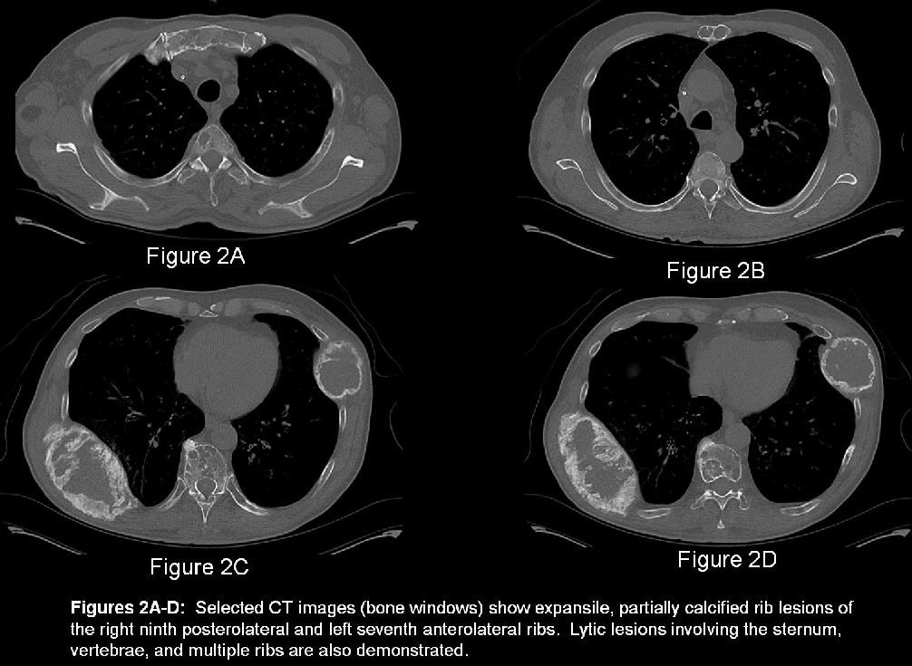
- Demonstrates many of the same features seen on radiography but to better advantage (Figure 2a-d)
- Marrow replacement by tumor cells
- Osseous lysis
- Trabecular and cortical destruction
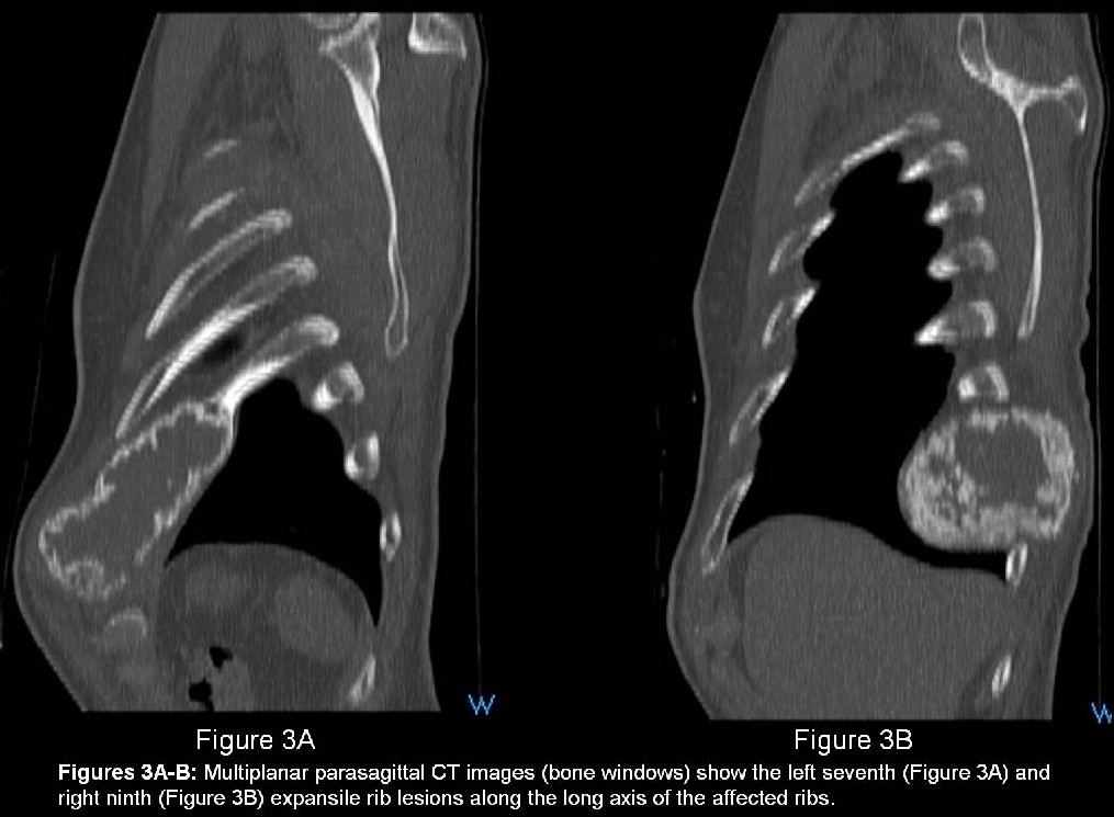
- Multiplanar (Figure 3a and 3b) and 3-D (Figure 4) imaging are helpful in delineating the relationship of the expansile lesions to various osseous structures
MRI
- Focal marrow replacement or diffuse marrow infiltration.
- Focal lesions are low signal intensity on T1WI, high signal intensity on T2WI.
- Following treatment lesions may demonstrate low signal intensity on T2WI.
Treatment
- Indications for treatment include
- Significant anemia
- Hypercalcemia
- Renal insufficiency
- Lytic bone lesions
- Extramedullary plasmacytomas
- Radiation Therapy
- Autologous Stem Cell Transplantation
- Chemotherapy
Prognosis
- Progressive course, median survival ~3 years
- 20-30% survive 5 or more years.
- <5% survive longer than ten years.
- Bone marrow labeling index and b2-microglobulin levels are most important prognostic indicators.
Caveats
- Skeletal survey preferred to scintigraphy.
- Multiple myeloma and metastatic disease would be the leading diagnostic considerations in patients over 50-years of age presenting with these imaging findings.
Suggested Readings:
- Burgener FA, Kormano M. Differential Diagnosis in Conventional Radiology, 2nd ed. New York: Thieme, 1991: 75-82.
- Guermazi A, Abdelwahab IF. Radiologic Imaging in Hematologic Malignancies. Birkhauser, 2004: 297-300.
- Lee JK, Sagel SS, Stanley RJ, Heiken JP. Computed Body Tomography with MRI Correlation. Lippincott Williams and Wilkins, 2005:622-625.
- Munk PL, Ryan A. Teaching Atlas of Musculoskeletal Imaging. New York: Thieme, 2007: 258-261.
- Cecil RL, Goldman L, Ausiello D. Cecil Textbook of Medicine, 22nd ed. Saunders, 2004: 1187-1192.
- Kumar V, Abbas AK, Fausto N. Robbins and Cotran Pathologic Basis of Disease, 7th ed. Elsevier Saunders, 2005: 679-681.
Original case written by its authors at Virginia Commonwealth University and published at this address as part of a weekly teaching collection. Reproduced here as an archive.