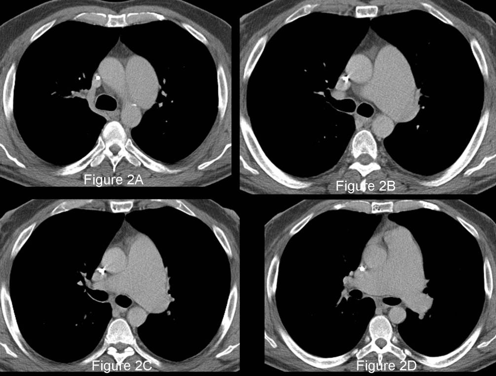
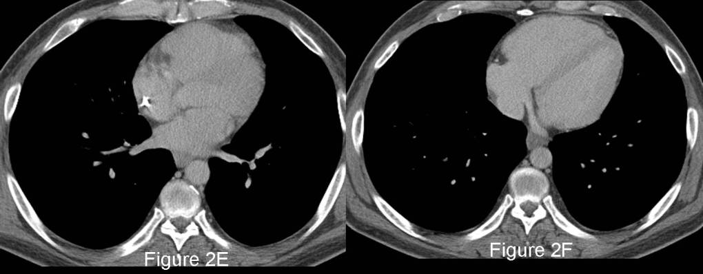
Radiologic Findings PA (Fig. 1A) and lateral (Fig. 1B) chest radiographs reveal marked enlargement of the main and left pulmonary artery. The right pulmonary artery is non-enlarged. The heart size is normal. Specifically, the right ventricle is not enlarged. The left thorax appears oligemic relatively to the right. The lungs are clear. A right upper extremity PICC line projects over the superior vena cava. Contrast-enhanced axial CT images through the main and central pulmonary arteries (Fig. 2A-D) reveal a suboptimal contrast bolus. The main pulmonary artery is enlarged (Fig. 2B) measured at 48 mm. The left pulmonary artery is also enlarged (Fig. 2C and 2D) measured at 37 mm. The right pulmonary artery is normal sized (Fig. 2C) measuring 17 mm. Note the absence of mediastinal and hilar lymphadenopathy. The myocardium and right ventricle in particular are not enlarged (Fig. 2E and 2F) (Images courtesy of John Grizzard, MD, VCU Medical Center, Cardio-Thoracic Imaging Section, Richmond, Virginia). Diagnosis: Pulmonic Stenosis
Differential Diagnosis
Discussion
Pulmonic stenosis results from a congenital malformation of the pulmonary valve and is the most common congenital anomaly that produces obstruction of the right ventricular outflow tract. The normally trileaflet valve may have fused commissures or may demonstrate a bicuspid or dysplastic morphology. It occurs as an isolated anomaly in up to 7% of patients with congenital heart disease. It is a common congenital cardiac lesion among those that initially manifest in adulthood.
Clinical Findings
Patients with pulmonic stenosis are acyanotic. Many patients are entirely asymptomatic, and most present in the third to fourth decades of life. Physical examination often reveals a harsh long (diamond-shaped) systolic ejection murmur. Patients with severe obstruction from pulmonic stenosis may present in childhood with easy fatigability and right ventricular failure.
Imaging Findings
Chest Radiography
- Mild to moderate main pulmonary artery enlargement (Fig. 1A and 1B)
- Left pulmonary artery enlargement (Fig. 1A and 1B)
- Normal right pulmonary artery, peripheral pulmonary arteries and pulmonary veins (Fig. 1A and 1B)
- Normal heart size (Fig. 1A and 1B)
- Rarely, pulmonic valve calcification
- Right ventricular enlargement in cases of right ventricular failure
CT/MRI
- Main and left pulmonary artery enlargement (Fig. 2A-2D)
- Demonstration of normal right pulmonary artery (Fig. 2C and 2D)
- Velocity encoded cine MR imaging; demonstration of differential blood flow in the pulmonary arteries and estimation of gradient across the stenotic valve
- Turbulent flow on gradient-echo cine sequences
- Normal heart size; eventual right ventricular hypertrophy from chronic pressure overload
Treatment
- Balloon dilatation for reduction of transvalvular gradient
- Repeat dilatation or open surgical correction (valve replacement) in approximately 20% of treated patients
Prognosis
- Good with appropriate treatment
- Survival beyond 50 years unusual in untreated patients
- Mortality and morbidity from infective endocarditis
Caveats
- Pulmonary artery enlargement in pulmonic stenosis results from post-stenotic dilatation secondary to the high velocity jet of blood forced through the stenotic valve However, the degree of dilatation is not related to the severity of the obstruction.
- Trilogy of Fallot refers to pulmonic stenosis complicated by right ventricular hypertrophy and a right-to-left shunt through an incompetent foramen ovale
- Pulmonic stenosis and pulmonary arterial hypertension may both manifest with pulmonary artery enlargement. The two conditions are differentiated by the fact that the right pulmonary artery is normal in pulmonic stenosis and enlarged in pulmonary arterial hypertension.
Selected Readings
- Gross GW, Steiner RM. Radiographic Manifestations of Congenital Heart Disease in the Adult Patient. Radiol Clin North Am 1991; 29: 293-317.
- Parker MS, de-Christenson Rosado ML, Abbott GF. Developmental Anomalies: Cardiac Anomalies-Pulmonic Stenosis. In: Teaching Atlas of Chest Imaging 2006; Thieme, New York;
- Steiner RM, Reddy GP, Flicker S. Congenital Cardiovascular Disease in the Adult Patient. Imaging update. J Thorac Imag 2002; 17: 1-17.
can you purchase viagra online where to purchase viagra trusted viagra sites generic ed medication buy amoxicillin cheap generic viagra online amoxicillin buy can you purchase viagra online where to purchase viagra trusted viagra sites generic ed medication buy amoxicillin cheap generic viagra online amoxicillin buy can you purchase viagra online where to purchase viagra trusted viagra sites generic ed medication buy amoxicillin cheap generic viagra online amoxicillin buy can you purchase viagra online where to purchase viagra trusted viagra sites generic ed medication buy amoxicillin cheap generic viagra online amoxicillin buy
Original case written by its authors at Virginia Commonwealth University and published at this address as part of a weekly teaching collection. Reproduced here as an archive.