Archived case 79 · Dec 10-Dec 17, 2010
Accessory Cardiac Bronchus
Clinical Presentation: Young patient involved in a high speed motor vehicle collision
The question posed to readers
What is the pertinent radiologic imaging finding?
Archived case 79 · Dec 10-Dec 17, 2010
Clinical Presentation: Young patient involved in a high speed motor vehicle collision
What is the pertinent radiologic imaging finding?
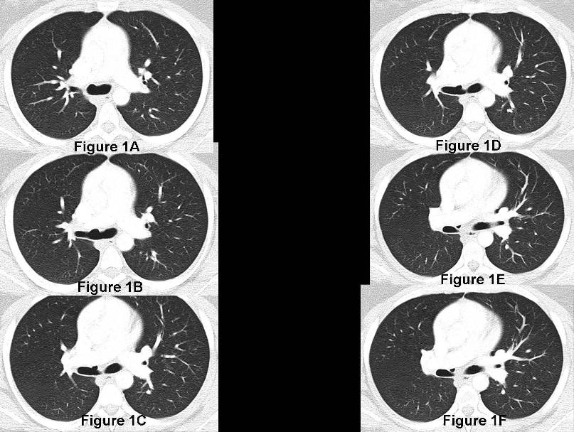
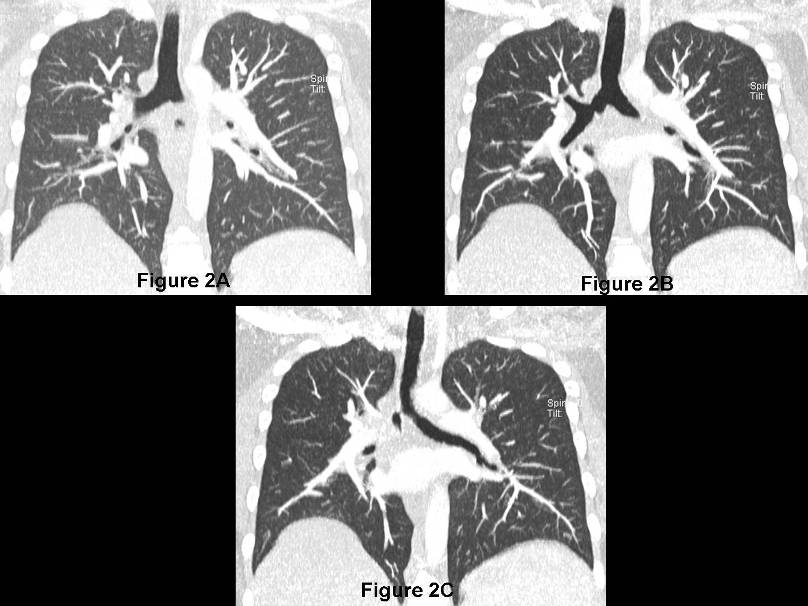
Accessory Cardiac Bronchus
Differential Diagnosis
None
Discussion
Variations of bronchial branching are common and may affect any portion of the tracheobronchial tree. These variations and are usually discovered incidentally. Right-sided anomalous bronchi are classified as pre-eparterial or post-eparterial if they arise proximal or distal to the origin of the right upper lobe bronchus respectively. Left-sided anomalous bronchi are classified as pre-hyparterial and post-hyparterial if they arise proximal or distal to the left upper lobe bronchus respectively. Upper lobe anomalous bronchi are much more common on the right than on the left. Displaced and supernumerary or coexistent normal upper lobe anomalous bronchi may also occur. Accessory cardiac bronchus is an anomalous bronchus that arises from the medial aspect of the right main stem or bronchus intermedius and courses towards the mediastinum. It is typically blind-ending but may be surrounded by lung parenchyma.
Clinical Findings
Most individuals with anomalous bronchi are asymptomatic and are diagnosed incidentally on chest CT. However, cases of hemoptysis and recurrent infection have been reported in association with anomalous bronchi.
Imaging Findings
Chest Radiography
MDCT
Treatment
Selected Readings
Filed under: Radiology, Medicine/Pulmonary
Original case written by its authors at Virginia Commonwealth University and published at this address as part of a weekly teaching collection. Reproduced here as an archive.