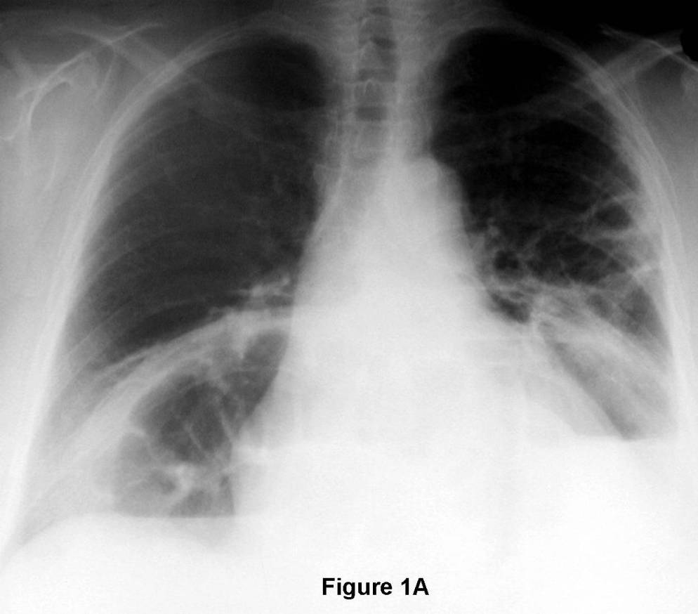
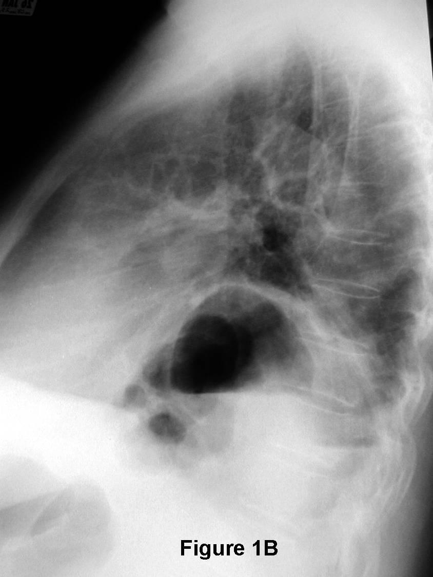
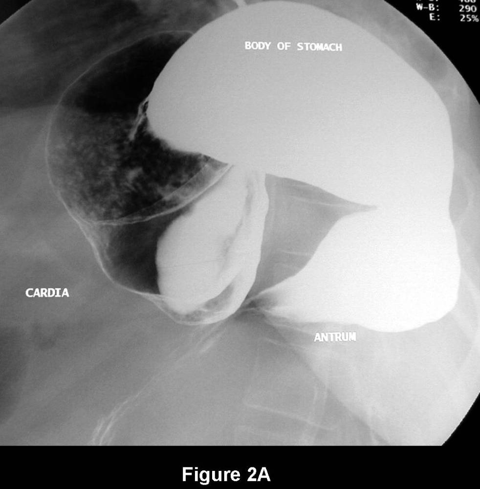
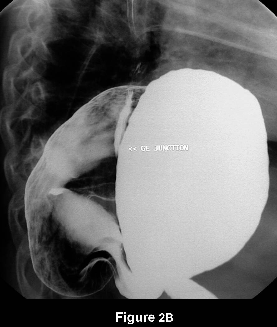
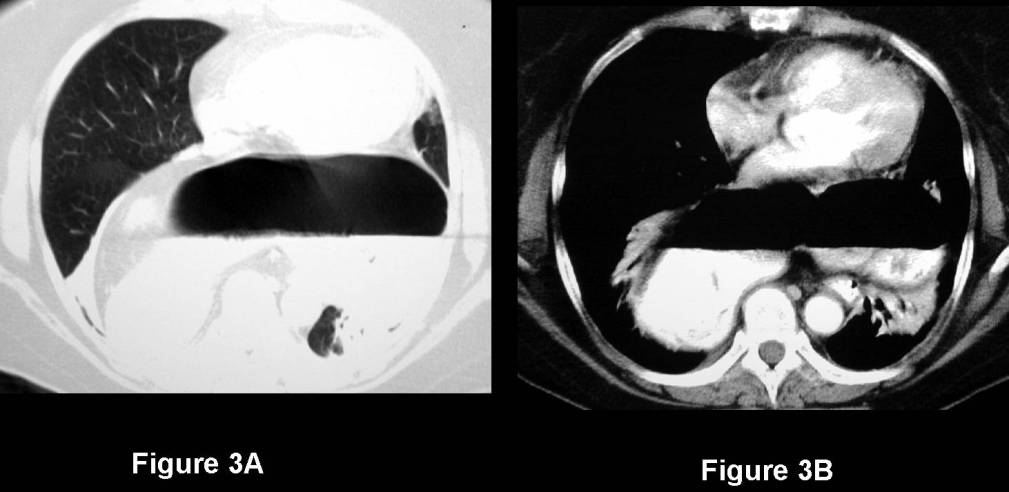
Radiologic Findings PA (Fig. 1A) and lateral (Fig. 1B) chest radiographs demonstrate an abnormal dilated curvilinear air-filled structure projected above the diaphragm and situated primarily behind the cardiac silhouette in the retrocardiac clear space (Fig. 1B). On closer inspection, this has the appearance of bowel and although haustral markings are suggested on the frontal exam (Fig. 1A) it appears featureless on the lateral exam (Fig. 1B). Additionally normal bowel is seen anteriorly below the diaphragm on the lateral exam (Fig. 1B) and subdiaphragmatic gastric fundal air is not seen. These results prompted a double contrast upper gastrointestinal series (Fig. 2A and 2B) which revealed a massive paraesophageal hernia with a total intrathoracic and upside-down stomach (i.e., volvulus). Note the position of the gastric cardia, gastroesophageal junction, and greater curvature of the stomach (Fig. 2A and 2B). These results are confirmed on the accompanying contrast-enhanced chest CT (Fig. 3A [lung windows] and Fig. 3B [mediastinal windows]). Note the absence of additional intrathoracic herniated hollow or solid subdiaphragmatic viscera. Diagnosis: Massive Type IV Paraesophageal Hernia and Gastric Volvulus
Differential Diagnosis
- Massive Sliding Hiatal Hernia
- Remote Diaphragmatic Rupture with Bowel Herniation
Discussion
There are two broad classifications of hiatal hernia. Sliding hiatal hernia by far accounts for the vast majority >90%) of cases. Paraesophageal hernias account for less than 5% of all hiatal hernias. As opposed to the sliding hiatal hernia, paraesophageal hernias are not associated with gastroesophageal reflux. Also, in contradistinction to the sliding hiatal hernia in which the gastroesophageal junction (GEJ) lies more than 2.0 cm above the diaphragm, paraesophageal hernias are characterized by a gastroesophageal junction (GEJ) located below the diaphragm. In this latter situation, the gastric fundus partially extends upwards through the esophageal hiatus and lies to the left of the distal esophagus. Some authors further divide hiatal hernias into four types. These types and their primary features are summarized in Table-1.
Hernia Type Classification Incidence Primary Characteristic I Classic Sliding >90% Widened esophageal hiatus allows stomach and possibly other intra-abdominal organs to prolapse into the thorax. GEJ is
Above diaphragmatic hiatus. Stomach forms posterior wall of the hernia sac.
II Pure Paraesophageal ~5% Part of the stomach (most commonly the fundus) herniates through the hiatus and lies alongside the distal esophagus. GEJ is
Below the diaphragm.
III Mixed ~5% Combined sliding and paraesophageal components. GEJ herniates into the thorax.
IV Massive (Total Intrathoracic Stomach) Rare Defect in central tendon of the diaphragm associated with slight transverse axis volvulus. Most or all of stomach resides in a huge intrathoracic peritoneal sac behind the heart. Other intra-abdominal organs often also herniate into the thorax (e.g. spleen, small Bowel).
In this particular case, the central tendon of the diaphragm was found to be completely disrupted at the time of exploratory laparotomy. It was postulated that the remote blunt thoracoabdominal trauma was responsible for this disruption which predisposed the patient to the eventual herniation.
Clinical Findings
Many patients with paraesophageal hernia are relatively asymptomatic. Others may complain of chest pain and dysphagia, or develop anemia. Strangulation and volvulus are unpredictable and may present with rapidly evolving and often fatal sepsis.
Complications of Paraesophageal Hernia
- Chronic / recurrent / asymptomatic occult GI blood loss → anemia (most common)
- Gastric volvulus (2nd most common)
Management
- Paraesophageal hiatal hernia should be surgically repaired if patient has no significant comorbid factors or contraindications to surgery
- Preoperative evaluation may include:
- Upper gastrointestinal series or Contrast-enhanced Chest and Abdominal CT
- Better define hernia type, anatomy, and additional intra-abdominal organs that may have herniated into the thorax
- Endoscopy
- Esophagitis
- Sricture
- Dysplasia
- Ulcerations
Selected Readings
- Schieman C, Grondin SC. Paraesophageal Hernia: Clinical Presentation, Evaluation, and Management Controversies. Thorac Surg Clin 2009; 19(40: 473-484.
- Kavic SM, Segan RD, George IM, et al. Classification of Hiatal Hernias Using Dynamic Three-Dimensional Reconstruction. Surg Innov 2006; 13(1): 49-52.
- Eren S, Ciris F. Diaphragmatic Hernia: Diagnostic Approaches with Review of the Literature. Eur J Radiol 2005; 54(3):448-459.
Original case written by its authors at Virginia Commonwealth University and published at this address as part of a weekly teaching collection. Reproduced here as an archive.