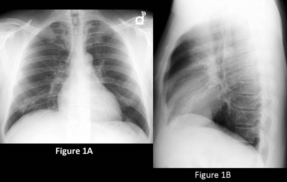
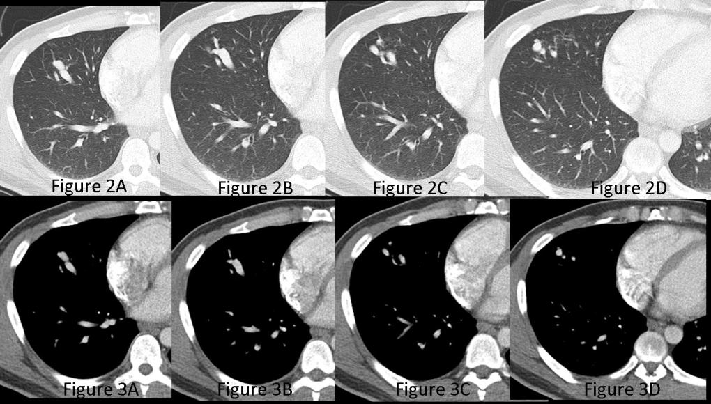
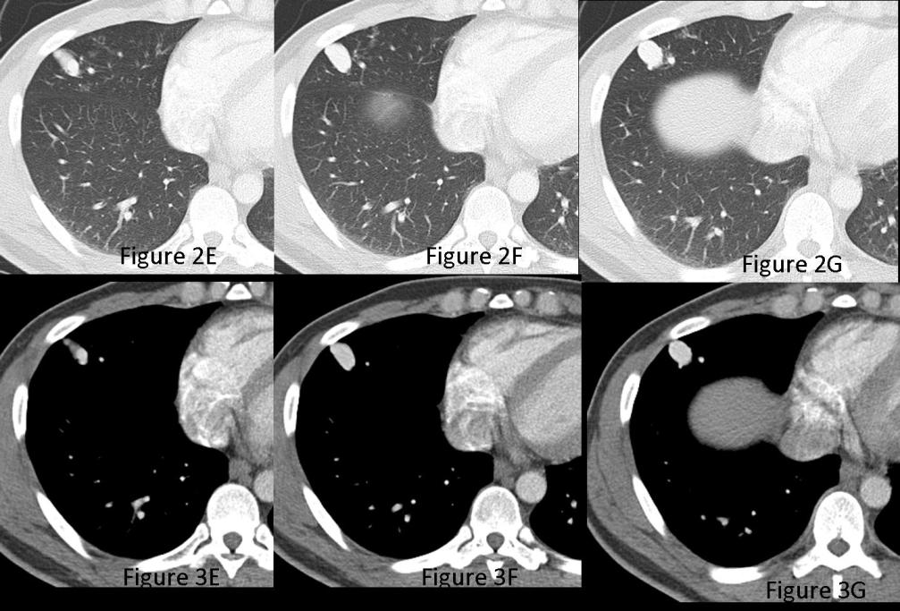
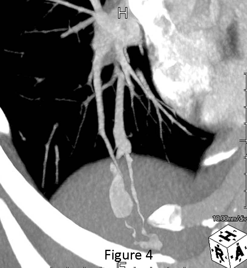
PA (Fig. 1A) and lateral (Fig. 1B) chest radiographs reveal an approximately 2.0cm lobulated non-calcified nodular lesion within the lateral segment of the right middle lobe. On closer inspection, the lesion is intimately related to and appears to be supplied by at least two enlarged blood vessels. Contrast-enhanced chest CT lung windows (Fig. 2A-G) with accompanying matched mediastinal windows (Fig. 3A-G) confirm the presence of an 18mm x 13mm enhancing lobulated lesion in the lateral segment of the right middle lobe. There is an enlarged feeding artery supplying the lesion and a dilated vein draining the lesion. The feeding artery and draining vein are nicely illustrated on the sagittal oblique CTA image (Fig. 4) (Images courtesy of Jennifer Hubert, MD, VCU Medical Center). Diagnosis: Pulmonary Arteriovenous Malformation
Differential Diagnosis
None
Discussion
A pulmonary arteriovenous malformation (PAVM) is an abnormal communication between a pulmonary artery and a pulmonary vein without an intervening capillary bed and results in a right-to-left shunt. Most AVMs likely stem from congenital defects in the capillary bed, which result in a direct communication between the pulmonary arterial and venous circulations. However, acquired arteriovenous communications may also occur following traumatic insults (non-iatrogenic or iatrogenic) or inflammatory process, and are usually designated arteriovenous fistulae. Osler-Weber-Rendu syndrome (aka hereditary hemorrhagic telangiectasia (HHT)) is an autosomal dominant disorder with a prevalence of one in 2,000 to 40,000 persons. Approximately 60 to 90% of PAVM occur in patients with HHT, up to 35% of patients with HHT have one or more PAVM, and 60% of patients with HTT-related PAVM have multiple lesions.
Clinical Findings
Over 50% of patients with PAVM are asymptomatic. Symptomatic patients are usually adults between the fourth and sixth decades of life. Symptoms may include fatigue and exertional dyspnea and neurologic complaints and/or fever from paradoxical emboli. Stroke occurs in up to 40% of patients with PAVM, brain abscess in 20%, and hemoptysis/hemothorax in 10%. Affected patients may have a family history of PAVM or HHT and may exhibit digital clubbing, cyanosis, and mucosal telangiectasias.
Imaging Findings
Radiography
- Lobular well-defined non-calcified nodule/mass (Fig. 1A; 1B)
- Most often located in peripheral lower lobe; often projects below the dome of the diaphragm
- Associated tortuous tubular opacities coursing to and from hilum representing feeding and draining vessels (Fig. 1A; 1B)
- Rarely multiple pulmonary nodules/masses
MDCT / CTA
- Nodular lesion or lesions with feeding artery(ies) and draining vein(s) (Fig. 2A-G; Fig. 3A-G; Fig. 4)
- Evaluate origin, number, length and diameter of feeding vessels and internal structure of vascular sac (Fig. 2A-G; Fig. 3A-G; Fig. 4)
- Rapid contrast enhancement and washout (Fig. 2A-G; Fig. 3A-G; Fig. 4)
- Unenhanced or enhanced 3D helical or multidetector CT for screening, characterization and quantification of PAVM (Fig. 2A-G; Fig. 3A-G; Fig. 4)
MRI / MRA
- Low signal flow-void in PAVM; low-to-intermediate signal in PAVM with internal thrombus
- 3-D MRA for non-invasive diagnosis of PAVM > 5 mm in size; high-signal intensity nodule and associated vessels
- Evaluate size and number of feeding vessels prior to embolotherapy
Angiography
- Opacification of feeding vessels and draining veins
- Confirm diagnosis, document presence of single or multiple lesions and evaluate origin, number, length and diameter of feeding vessels for coil embolization therapy
Treatment
- PAVM with feeding vessels larger than 3mm in diameter are traditionally treated with embolotherapy. However, a significant number of patients with smaller lesions suffer from clinically occult stroke. Thus, embolotherapy is often performed on all PAVM that can be super-selectively cannulated.
- Embolotherapy with detachable coils or occasionally detachable balloons for improvement of oxygenation and prevention of neurologic complications
Prognosis
- Significant morbidity and reported mortality rate of 10% for HHT patients
- Complications of embolotherapy may include: paradoxical emboli, air emboli, transient ischemic attacks, angina, bradycardia, hypotension, and chest pain
- Complete obliteration of right-to-left shunt: achieved <50% of treated patients
Selected Readings
- Coley SC, Jackson JE. Pulmonary arteriovenous malformations. Clin Radiol 1998; 53:396-404.
- Maki DD, Siegelman ES, Roberts DA, et al. Pulmonary arteriovenous malformations: three-dimensional gadolinium-enhanced MR angiography-initial experience. Radiology 2001; 219: 243-246.
- Parker MS, Rosado-de-Christenson ML, Abbott GF. Developmental Anomalies: Anomalies of the Pulmonary Arteries and Veins. In: Teaching Atlas of Chest Imaging 2006; Thieme, New York, pp. 71-75.
- Pugash RA. Pulmonary arteriovenous malformations: overview and transcatheter embolotherapy. Can Assoc Radiol J 2001; 52: 92-102.
Original case written by its authors at Virginia Commonwealth University and published at this address as part of a weekly teaching collection. Reproduced here as an archive.