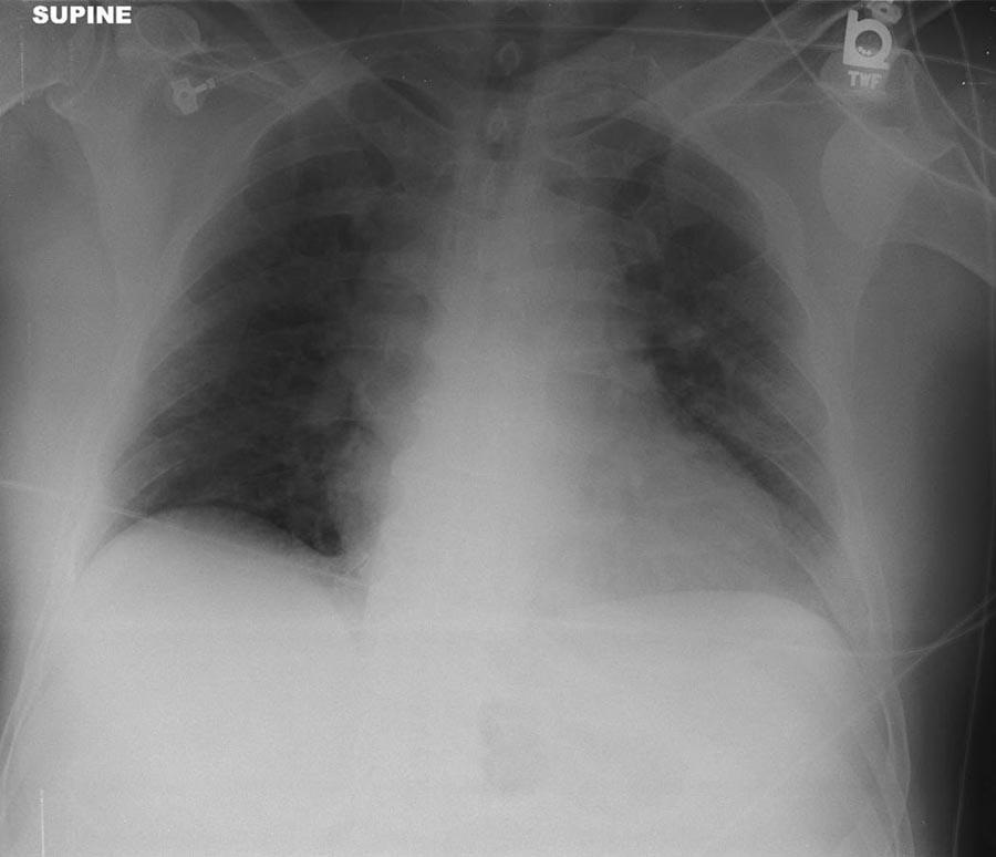
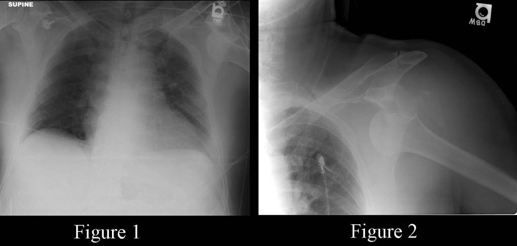
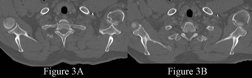
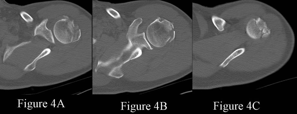
AP supine chest radiograph obtained in the trauma bay (Fig. 1) demonstrates an anterior inferior dislocation-fracture of the left glenohumeral joint with associated traumatic Hill-Sachs deformity. There is slight widening of the mediastinum and an indistinct right superior mediastinal border. Mediastinal hematoma and potential aortic injury cannot be excluded on the basis of this exam alone. Additionally, there is an acute left 7th rib fracture and ground glass opacity in the left lower lobe from pulmonary contusion. Dedicated AP left shoulder radiograph (Fig. 2) confirms the anterior-inferior glenohumeral dislocation. The humeral head is located inferior and medial to the glenoid. Note the acute fracture fragments lateral to the glenoid. Selected axial images from the chest CTA (bone windows) (Fig. 3A and Fig. 3B) at the thoracic inlet demonstrate the acute fracture-dislocation to better advantage as well as the traumatic Hill-Sachs deformity. No acute thoracic aorta injury or mediastinal hematoma was identified on the CTA. Unenhanced axial CT images of the left shoulder obtained following closed reduction (Fig. 4A-C) reveal anatomic reduction of the left glenohumeral joint. The proximal humeral fracture is more fully characterized, revealing a comminuted minimally displaced fracture involving both the lesser and greater tuberosity. Diagnosis: Acute Left Glenohumeral Fracture-Dislocation with Traumatic Hill-Sachs Lesion
Differential Diagnosis
None
Discussion
Portable chest radiography is commonly used in the Trauma bay by Emergency Room Physicians, Trauma Surgeons, and Radiologists, as a diagnostic adjunct in the primary survey of trauma patients, and to evaluate for potentially life-threatening and otherwise unsuspected complications (e.g., pneumothorax, hemothorax, diaphragmatic injury, possible mediastinal injury, etc). Additional unsuspected concomitant non-life-threatening but significant injuries may also be detected by the astute radiologist (e.g., Thoracic spine injuries; scapular fractures; rib fractures; sternoclavicular dislocation; acromion clavicular disruption; clavicle fractures; glenohumeral fracture-dislocations, etc). Such injuries may otherwise go undetected or unnoticed in the polytrauma victim with more severe injuries demanding more emergent attention or in the neurologically altered or impaired patient (e.g., head injury,; drug and or alcohol intoxication) resulting in increased morbidity and possible disability.
Etiology
Glenohumeral Dislocation
Anterior Dislocation
- 10X more common; accounts for 95% of shoulder dislocations
- Mechanism of injury: Usually traumatic, most commonly occurs when the affected arm is forcibly externally rotated and abducted (e.g., when football players arm tackle or skiers planted uphill pole gets stuck); less often results from a direct blow to the back of the shoulder
Imaging Findings
- Diagnosis easily made on AP chest radiography if the shoulder is included in field-of-view or on dedicated AP shoulder radiography
- Humeral head lies anterior, inferior, and medial to glenoid
- Associated injuries
- Hill Sachs lesion: humeral head impacts against inferior lip of glenoid, causing an indentation posteriorly and superiorly
- Bankart lesion: avulsion or bony fragment off the inferior glenoid; less common
Posterior Dislocation
- Less common injury; 5% shoulder dislocations
- More difficult clinical and radiologic diagnosis
- Mechanism of injury: May involve forceful muscle contraction (tonic-clonic seizures, electrocution), fall on a flexed, internally rotated, and adducted arm, or direct forceful blow to the anterior shoulder
Imaging Findings
- Frontal AP views may appear normal; subtle clue may be the absence of the normal “crescent” sign caused by bony overlap of the normal humeral head and glenoid
- “Fixed” in internal rotation
- Associated injuries
- “Trough” sign-15% injuries; compression fracture of anterior humeral surface
- “Reverse Bankart lesion”-disruption of posterior rim of glenoid
Transscapular view
- Best conventional radiographic method to unequivocally diagnosis shoulder dislocation
- Provides en face view of the glenoid and depicts the relationship of the humeral head to the glenoid itself (i.e., normal, anterior, posterior)
MDCT
- Provides confirmatory data
- Additional information with respect to concomitant fractures and intra-articular bony fragments; intramuscular hematoma; and hemoarthrosis
Selected Readings
- Chew FS, Bui-Mansfield LT, Kline MJ. Trauma in Adults: Upper Extremity In: Musculoskeletal Imaging 2003; Lippincott Williams & Wilkins, Philadelphia: 35 – 77.
- Ho ML, Gutierrez FR. Chest Radiography in Thoracic Polytrauma. AJR 2009; 192: 599-612.
- Manaster BJ, Disler DG, May DA. Upper Extremity Trauma In: Musculoskeletal Imaging: The Requisites 2002; Mosby, St. Louis: 209-250.
Original case written by its authors at Virginia Commonwealth University and published at this address as part of a weekly teaching collection. Reproduced here as an archive.