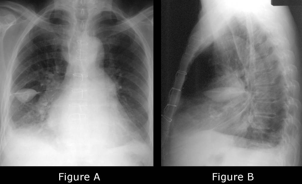
Radiologic Findings PA (Fig. A) and lateral (Fig. B) chest radiographs demonstrate evidence of a remote median sternotomy. The cardiomediastinal silhouette is enlarged and vascular redistribution is evident. A right-sided pleural effusion is present which partially silhouettes the ipsilateral diaphragm and blunts the posterior and lateral sulcus. An ovoid radio-opacity is present in the right hemithorax. The long axis of this opacity parallels the long axis of the horizontal fissure. The opacity also changes its morphology from one orthogonal view to the next, relatively foreshortened on the frontal exam (Fig. A) and becoming more elongated on the lateral exam (Fig. B). Both of the latter findings confirm the lesion in question is pleural-based and not parenchymal-based. Also note the absence of air-bronchograms. Diagnosis: Interlobar Pseudotumor; Cardiac Decompensation
Differential Diagnosis
- Hematoma
- Pleural Metastases
- Primary Pleural Neoplasms
Discussion
Interlobar Pseudotumor is known by various terms including interlobar fluid, interfissural fluid, pseudotumor, phantom tumor, and vanishing tumor. For unknown reasons, the pleural effusion associated with heart failure may occasionally localize in one of the interlobar fissures simulating a mass or true tumor. Although this may occur in any standard or accessory fissure, there is a predilection for such to occur in the horizontal fissure of the right lung (78% cases). Interlobar fluid collections may also be seen less frequently in the setting of renal failure and hepatic hydrothorax. An important point to remember is that these fluid collections are simply “localized” and not “loculated” within the fissure. A point easily illustrated with dependent decubitus radiographs which can be acquired in problematic or questionable cases.
Clinical Findings
The clinical findings are related to the underlying responsible disease process, most often heart failure, and not the pleural effusion itself.
Imaging Findings
- Homogeneous ovoid opacity oriented along long axis of fissure (Fig A and B)
- Evidence of ipsilateral pleural fluid (Fig A and B)
- Absence of air-bronchograms (Fig A and B)
- Morphology changes from one orthogonal view to the next (Fig A and B)
- Mobile with decubitus positioning
- Invariably additional radiographic evidence of concomitant or resolving heart failure (Fig A and B)
Treatment
- Directed toward the underlying disease process
Prognosis
- Related to the underlying disease process
- Interlobar fluid collections tend to resorb spontaneously, hence the alternate designations “phantom tumor” and “vanishing tumor”
Selected Readings
- Fraser RS, Müller NL, Colman N, Paré PD. Pleural Abnormalities. In: Fraser and Pare’s Diagnosis of Diseases of the Chest, 4th ed. W.B. Saunders Company, Philadelphia 1999; 579-581.
- Haus BM, Stark P, Shofer SL, Kuschner WG. Massive Pulmonary Pseudotumor. Chest 2003; 124(2):758-760.
Original case written by its authors at Virginia Commonwealth University and published at this address as part of a weekly teaching collection. Reproduced here as an archive.