Archived case 6 · Jul 2-Jul 9, 2009
Elastofibroma dorsi
A 79-year-old man complains of a “clicking” sensation in his left shoulder with activity and has a palpable infrascapular mass.
Archived case 6 · Jul 2-Jul 9, 2009
A 79-year-old man complains of a “clicking” sensation in his left shoulder with activity and has a palpable infrascapular mass.
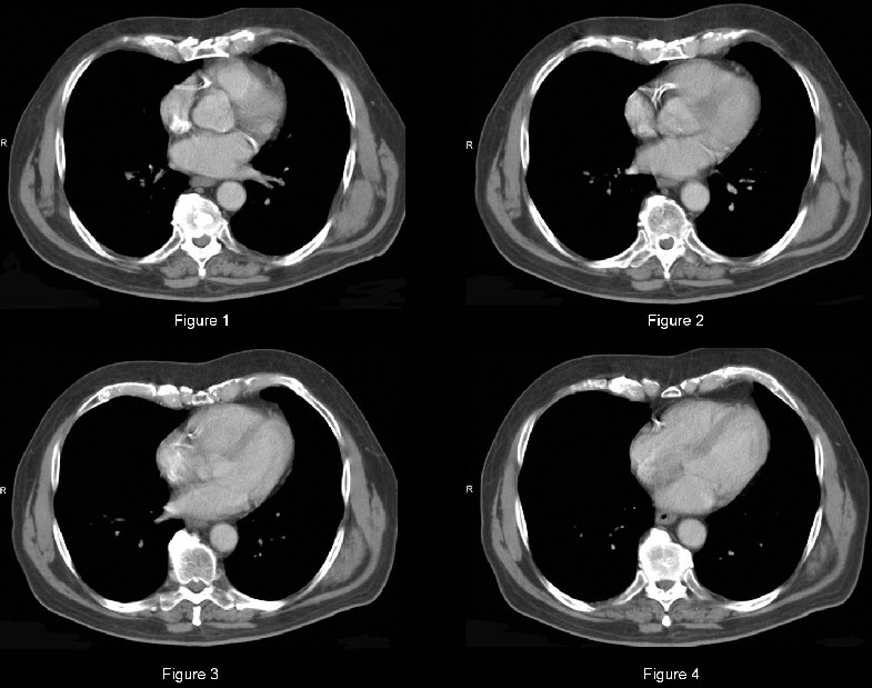
Elastofibroma dorsi
Differential Diagnosis
Infrascapular lesion of decreased-to-intermediate attenuation similar to skeletal muscle:
· Extrabdominal desmoid
· Neurofibroma
· Cicatricial fibroma
· Malignant fibrous histiocytoma
· Sarcoma
Discussion
Background
Elastofibroma dorsi (ED) is a slow growing, benign lesion of unknown cause and is most commonly seen in the periscapular or infrascapular regions of the chest wall. Autopsy series report ED 3 cm or smaller in 24% of women and 11% of men more than 55 years old. The name reflects its characteristic location at the medial, inferior border of the scapula. ED lesions are periscapular in 99% of cases and are bilateral in 10-66% of cases. Synchronous lesions in the infraolecranon region are also common. Other reported locations include the thoracic wall, deltoid muscle, axilla, ischial tuberosity and greater trochanteric region.
Etiology
Although the etiology is unclear, there is an increased prevalence of ED in manual laborers. Thus, it has been postulated that ED may be reactive in nature and attributable to repetitive mechanical friction of the scapula against the ribs. This theory provides an explanation for the right-sided preponderance. However, up to 32% of cases occur in patients with a family history of ED, suggesting a genetic, nontraumatic origin.
Clinical Findings
Elastofibromas occur most often in elderly women but have been reported in persons aged 6-94 years. Most patients with ED are asymptomatic and small pseudotumors may be overlooked unless the patient is asked to move his or her arm laterally or anteriorly. Only rarely do patients report stiff shoulders, local pain with arm movement, and/or an annoying “click” when using their shoulder.
Imaging Findings
Radiography
· Usually normal
Ultrasonography
One of four ultrasound patterns may be detected:
· Type I (54%): inhomogeneous fasciculated.
· Type II (22%): inhomogeneous nonspecific.
· Type III (15%): hyperechogeneous.
· Type IV (9%): hypoechogeneous.
· In those cases without a clearly defined cleavage plane, ED is difficult to differentiate from surrounding
muscle.
CT
· Poorly defined, inhomogeneous, unencapsulated, lenticular, soft-tissue mass.
· Attenuation similar to that of skeletal muscle.
· Contains linear streaks of fat attenuation.
MRI
· Similar appearance to CT.
· T1 / T2-weighted images: Signal intensity similar to that of skeletal muscle with interlaced streaks of fat signal intensity.
· Heterogenous enhancement after gadolinium administration.
Treatment
· Asymptomatic patients: No treatment necessary.
· Symptomatic patients:
Prognosis
· Recurrence after surgery is unusual.
· No cases of malignant transformation have been reported.
Caveats
· In most cases, the diagnosis can be made on the basis of the characteristic CT /MRI imaging findings and the typical periscapular or infrascapular anatomic location, avoiding unnecessary biopsy and surgery in asymptomatic patients.
· Elastofibromas are very frequently bilateral. The presence of a similar contralateral periscapular lesion is of great benefit in establishing the correct diagnosis, virtually eliminating malignancy from the differential.
Suggested Readings
1. Battaglia M, Vanel D, Pollastri P, Balladelli A, Alberghini M, Staals EL, Monti C, Galletti S. Imaging patterns in elastofibroma dorsi. Eur J Radiol June 16. [Epub ahead of print].2. Kransdorf MJ, Meis JM, Montgomery E. Elastofibroma: MR and CT appearance with radiologic-pathologic correlation. AJR 1992; 159:575-579.
3. Naylor MF, Nasciemento AG, Sherrick AD, McLeod RA. Elastofibroma dorsi: Radiologic findings in 12 patients. AJR 1996; 167:683-687.
Filed under: Radiology, Medicine/Pulmonary
Original case written by its authors at Virginia Commonwealth University and published at this address as part of a weekly teaching collection. Reproduced here as an archive.