Thoracic Imaging Archive

Archived case 58 · Jul 30-Aug 6, 2010

Asbestos-Related Pleural Plaques

Clinical Presentation: Withheld

The question posed to readers

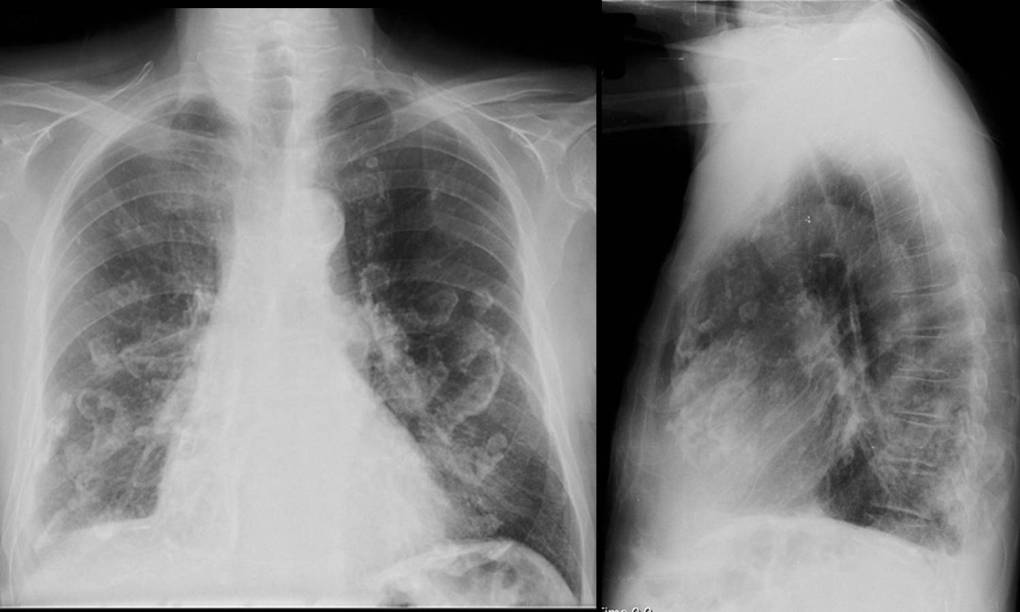
What are the pertinent radiologic findings? What is your leading diagnosis?
Images

Radiograph 1 from archived case 58
Figure 1

Diagnosis

Asbestos-Related Pleural Plaques

65-year-old asymptomatic man who had been employed in construction and demolition for over forty years (originally withheld) Radiologic Findings PA (Figure A) and lateral (Figure B) chest radiographs demonstrate the presence of bilateral, relatively symmetric, multi-focal, discontinuous areas of pleural thickening and calcification primarily distributed along the anterolateral and posterolateral chest wall and domes of each hemidiaphragm. The apices and costophrenic angles are spared. Those lesions seen en face on the frontal exam (Figure A) exhibit scalloped or “holly-leaf” morphology, whereas those seen in profile on the lateral exam (Figure B) appear more linear confirming the lesions change morphology from one orthogonal plane to the next and are therefore pleural-based.     Diagnosis: Asbestos-Related Pleural Plaques

Differential Diagnosis

None

Discussion

Asbestos-related “plaques” are a pleural-based process that MUST be differentiated from “asbestosis” which is a parenchymal-based disease process. Although the two processes may be seen in the same patient, they are two different types of lesions and interchanging the two terms is incorrect. There are five pleural and three pulmonary parenchymal manifestations of asbestos exposure as listed the Table provided below:

  Pleural-Based Manifestations of                      Asbestos Exposure Parenchymal-Based Manifestations of  A sbestos Exposure Benign Exudative Pleural Effusion Asbestosis Diffuse Pleural Thickening Rounded Atelectasis Non-calcified Pleural Plaques Primary Lung Cancer Calcified Pleural Plaques   Diffuse Malignant Mesothelioma  


Pleural plaques are the macroscopic and radiologic hallmarks of past asbestos exposure and typically develop 15-20 years following the initial exposure. Pleural plaques involve the parietal pleura and tend to occur adjacent to relatively rigid structures such as the ribs, vertebrae, and central tendon of the diaphragm. Subsequent calcium deposition often occurs in the pleural plaques. These initially appear as fine, punctuate flecks and then often coalesce over time to form dense streaks or plate-like deposits.


Clinical Findings

Patients with asbestos-related pleural plaques are usually asymptomatic. Patients with asbestosis are usually symptomatic and often experience dyspnea, dyspnea on exertion, cough, hypoxia, and restrictive physiology on pulmonary function studies (PFTs).


Imaging Findings

  • Bilateral, discontinuous, multi-focal areas of pleural thickening (Fig. A and Fig. B)
  • Located along posterolateral chest wall between 7th-10th ribs and lateral chest wall between the 6th-9th ribs (Fig. A and Fig. B)
  • May involve the mediastinal pleura and tendinous insertions of diaphragm (Fig. A and Fig. B)
  • Usually spares apices, interlobar fissures, costophrenic angles, and visceral pleura (Fig. A and Fig. B)
  • Variable degrees and patterns of calcification (Fig. A and Fig. B)
  • Serpentine marginal calcification creates a “holly-leaf” pattern when viewed en face (Fig. A and Fig. B)
  • Morphologic pattern or appearance of pleural plaques changes when viewed in profile versus that seen en face (Fig. A and Fig. B)

Treatment

  • None indicated

Prognosis

No proven long-term sequelae from the plaques alone


Caveats

  • “Asbestosis” SHOULD NOT be confused with “asbestos-related pleural disease”. The former is an interstitial lung disease characterized by pulmonary fibrosis resulting from exposure to “asbestos”.  The latter does not affect the lung but the pleura and typically occurs in asymptomatic persons.
  • Persons are NOT exposed to “asbestosis” but rather have various occupational exposures to “asbestos”. “Asbestosis” in a parenchymal disease and NOT something to which you can be exposed.

Selected Readings

  1. Parker MS, Rosado-de-Christenson ML, Abbott GF. Asbestos-Related Pleural Disease. In: Teaching Atlas of Chest Imaging. Thieme, New York: 2006; 716-720.
  2. Parker MS, Rosado-de-Christenson ML, Abbott GF. Asbestosis. In: Teaching Atlas of Chest Imaging. Thieme, New York: 2006; 523-526.

Filed under: Radiology, Medicine/Pulmonary

Original case written by its authors at Virginia Commonwealth University and published at this address as part of a weekly teaching collection. Reproduced here as an archive.

Back to the case index