Aortic Coarctation; Severe High Grade Stenosis and Extensive Collateral Flow to the Descending Thoracic Aorta
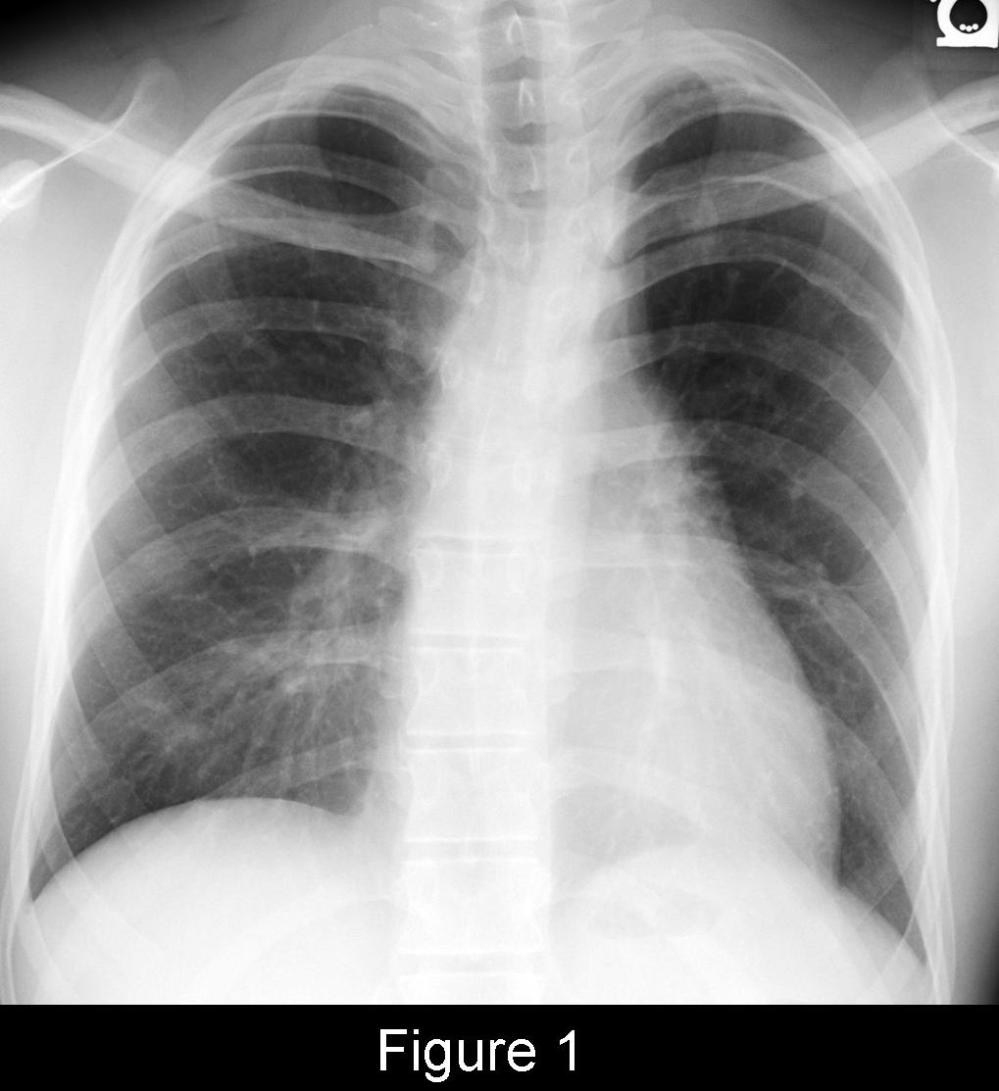
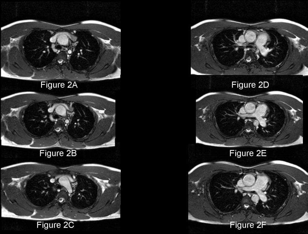
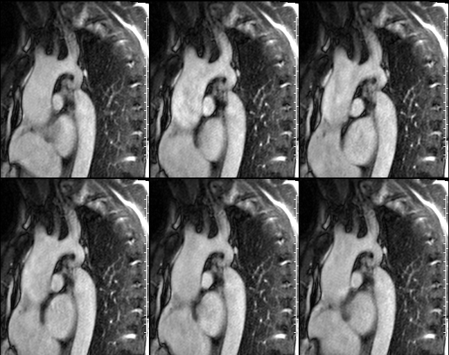
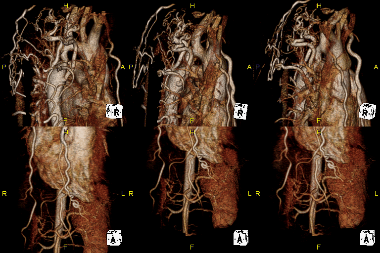
Clinical Presentation: 22-year-old man presenting with fatigue, claudication, and differential blood flow between the upper and lower extremities (originally withheld) Radiologic Findings PA chest radiograph (Fig. 1) demonstrates a small or inconspicuous transverse aorta with a morphology that resembles the number 3 (“aortic 3” sign). The heart size and pulmonary vascular clarity are normal. There is also severe rib notching of the inferior borders of the 3rd through 6th ribs bilaterally. Selected sequential cardiac MRI FISP axial (Fig. 2A-2F) and sagittal oblique FISP (Fig. 3) images reveal a severe high grade narrowing of the thoracic aorta, just distal to the origin of the left subclavian artery. Flow studies obtained in the plane of the aortic arch (not illustrated) demonstrated trace flow traversing the region. Color 3-D volume rendered MRA (Fig. 4) shows numerous dilated intercostal and internal mammary collateral vessels extending to the descending aorta (Images courtesy of Jennifer Hubert, MD, and John Grizzard, MD, VCU Medical Center) Diagnosis: Aortic Coarctation; Severe High Grade Stenosis and Extensive Collateral Flow to the Descending Thoracic Aorta
Differential Diagnosis
- Neurofibromatosis
- Pulmonary Atresia with Harvested Systemic Vessels Communicating with Pulmonary Vessels
Discussion
Coarctation is a congenital anomaly of unknown etiology that results in a focal obstruction at the junction of the distal aortic arch and descending thoracic aorta. It is responsible for 6% of congenital cardiac anomalies. Although there is blood flow across the coarc segment, it is supplemented by collateral blood flow that bypasses the obstruction via intercostal, internal mammary, epigastric, spinal, and periscapular arteries. Rib notching is created by pulsation of dilated intercostal arteries providing the collateral blood flow. It is rare before 10-years of age and is most pronounced along the posterior upper ribs. The 1st and 2nd rib are usually spared because of communication between the interccstal arteries and aorta above the coarctation. The”figure-of -3 –sign” results from dilatation of the left subclavian artery because of increased pressures proximal to the coarctation and the post-stenotic dilatation of the descending aorta.
Clinical Findings
Isolated coarctation more commonly occurs in men than in women (2.5:1 ratio). Coarctation associated with congenital heart disease affects men and women equally. 20% of women with Coarctation also have Turner syndrome. Other associated findings include: bicuspid aortic valve (50%); mitral valve prolapse; patent ductus arteriosus; and ventricular septal defects. Those patients with aortic coarctation and associated anomalies typically are symptomatic at birth, often presenting in heart failure. Patients with isolated coarctation may be asymptomatic or may become symptomatic in childhood or young adulthood, and often complain of fatigue, claudication, and headache. Alternatively, such affected patients may present with systemic hypertension, valvular murmur, endocarditis, and aortic dissection. Physical examination often reveals differential blood flow between the upper and lower extremities.
Imaging Findings
Chest Radiography (Children and Young Adults)
- Normal heart size and pulmonary vascular clarity (Fig. 1)
- Cardiomegaly from left ventricular hypertrophy and left ventricular failure
- Narrow cardiac waist (Fig. 1)
- Small or inconspicuous aortic arch (Fig. 1)
- Figure-of-3-configuration of the lateral border of the aortic arch from post-stenotic dilatation distal to the coarctation (Fig. 1)
- Inferior rib notching; usually of ribs 3-9 (Fig. 1)
MDCT
- Visualize length and location of the coarctation and relative size of aorta proximal and distal to the coarc segment
- Visualize aortic branches and relationship of such to the coarctation
- Visualize collateral vessels
MRI
- Visualize and measure length and location of obstruction and degree of luminal compromise (Fig. 2-4)
- Visualize aortic branches and their relationship to the coarctation (Fig. 4)
- Visualize collateral vessels (Fig. 4)
- Velocity-encoded cine MRI sequences useful to estimate flow gradients across the coarctation
Treatment
- Resection of affected segment; graft placement
- Poor surgical candidates; balloon dilatation of the coarc segment may be beneficial
Prognosis
- Good with early surgical intervention
- Poor without surgical intervention; very high mortality rate by 6th decade of life in this subset of patients
Selected Readings
- Cole TJ, Henry DA, Jolles H, Proto AV. Normal and Abnormal Vascular Structures that Simulate Neoplasms on Chest Radiographs: Clues to Diagnosis. RadioGraphics 1995; 15: 867-891.
- Parker MS, Rosado-de-Christenson ML, Abbott GF. Coarctation of the Aorta. In: Teaching Atlas of Chest Imaging. Thieme, New York; 2006: 53-57.
- Steiner RM, Reddy GP, Flicker S. Congenital Cardiovascular Disease in the Adult Patient: Imaging Update. J Thorac Imaging 2002; 17: 1-17.
Original case written by its authors at Virginia Commonwealth University and published at this address as part of a weekly teaching collection. Reproduced here as an archive.