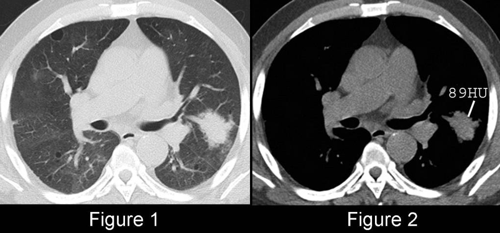
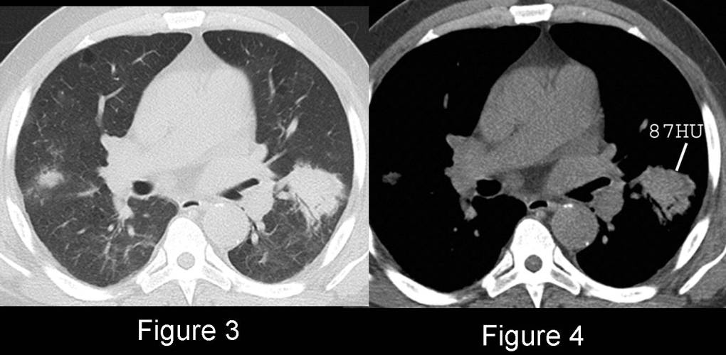
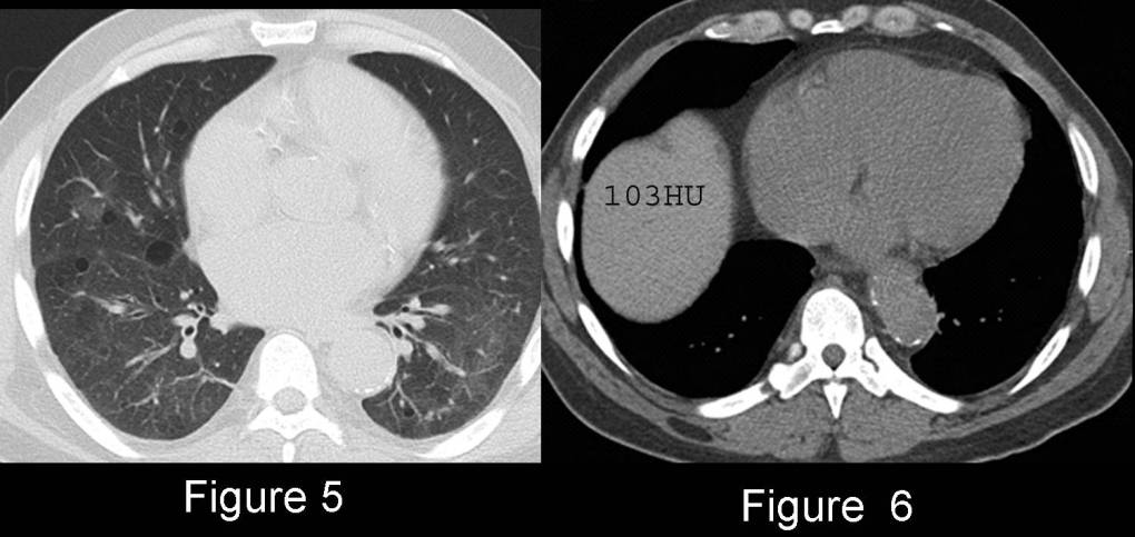
Unenhanced chest CT (lung windows) (Fig. 1; 3; 5) demonstrates patchy ground-glass opacities in both lungs and focal mass-like regions of consolidation in the lingula, left lower lobe, and lateral segment right middle lobe. Corresponding mediastinal windows (Fig. 2; 4) reveal the focal areas of consolidation are higher in attenuation (89HU and 87HU, respectively) than the soft tissues (38HU). Unenhanced CT through the liver (Fig. 6) shows it is also higher in attenuation than normal. Diagnosis: Amiodarone Pulmonary Toxicity
Differential Diagnosis
- Cryptogenic Organizing Pneumonia (COP)
- Pulmonary Fungal Disease
- Primary and Secondary Lung Neoplasia
Discussion
Background
Amiodarone is a tri-iodinated benzofuran derivative used to treat refractory tachyarrhythmias. Amiodarone contains 37% iodine by weight, has a long half-life (58-days), accumulates in the lung, and may cause a form of drug-induced lung injury that develops acutely, within a few days of initial use, or more than 10-years latter, while on chronic therapy. The precise incidence of pulmonary toxicity is unknown but it is estimated to occur in 5-7% of patients. Risk factors for pulmonary toxicity include: high cumulative dose, more than 400 mg/day, duration of use, increased age, and preexisting lung disease. Amiodarone also accumulates in the liver, spleen, muscles, adipose tissue, and skin.
Clinical Findings
Two distinct clinical presentations of pulmonary toxicity have been described. The most common is characterized by the insidious onset of dyspnea on exertion, dry cough, weight loss, weakness, and sometimes fever. Affetced patients most often exhibit an interstitial pattern of disease on imaging studies. One-third of patients have a more acute presentation that may mimic infection, pulmonary edema, or thromboembolism clinically and radiographically. The most specific test of Amiodarone pulmonary toxicity is a dramatically decreased diffusing capacity for carbon monoxide (DLCO) on PFT’s. Manifestations of extrapulmonary toxicity include: blue-gray skin discoloration, vortex corneal keratopathy, thyroid dysfunction, hepatic dysfunction, muscle weakness, and peripheral neuropathy.
Imaging Findings
Chest Radiography
- Diffuse bilateral reticular opacities
- Bilateral areas of consolidation; peripheral and predominantly upper lobes
- Focal consolidations and nodules; less common
- +/- pleural thickening
- Pleural effusion; uncommon
CT
- Ground-glass opacities (Fig. 1; 3; 5)
- Focal or diffuse areas of consolidation
- Reticular opacities
- Conglomerate mass(es); less common (Fig. 1-4)
- Consolidations / masses contain foci of attenuation greater than that of soft-tissue (82-175HU) (Fig. 1-4)
- Increased attenuation of liver or spleen and myocardium (Fig. 6)
Management
- Cessation of Amiodarone therapy
- Corticosteroids
Prognosis
- Usually good response to drug cessation and corticosteroids
- 5-10% of patients that develop pulmonary toxicity die as a sequelae
Caveats
- Increased liver attenuation on CT is also present in patients treated with Amiodarone without pulmonary toxicity
Selected Readings
- Camus P, Martin WJ, Rosenow EC. Amiodarone Pulmonary Toxicity. Clin Chest Med 2004; 25(1):65-75. Review.
- Papiris SA, Triantafillidou C, Kolilekas L, Markoulaki D, Manali ED. Amiodarone: Review of Pulmonary Effects and Toxicity. Drug Saf 2010; 33(7):539-558.
- Poll LW, May P, Koch JA, et al. HRCT Findings of Amiodarone Pulmonary Toxicity: Clinical and Radiologic Regression. J Cardiovasc Pharmacol Ther 2001; 6(3):307-311.
Original case written by its authors at Virginia Commonwealth University and published at this address as part of a weekly teaching collection. Reproduced here as an archive.