Archived case 5 · Jun 25-Jul 2, 2009
Empyema Thoracis
A 72-year-old woman presented to the emergency department with fever, cough, chest pain and shortness of breath.
Archived case 5 · Jun 25-Jul 2, 2009
A 72-year-old woman presented to the emergency department with fever, cough, chest pain and shortness of breath.
Empyema Thoracis
Differential Diagnosis
Lung abscess
Discussion
Background
Parapneumonic effusions (PPE) account for about one-third of all pleural effusions and are the most common cause of exudative effusion. Untreated or inadequately treated PPE may progress to empyema thoracis, which, by definition, is pus in the pleural space. The evolution of PPE is divided into three stages that represent a continuous spectrum. Stage I (exudative) (uncomplicated PPE): rapid outpouring of fluid into the pleural space due to increased pulmonary interstitial fluid traversing the pleura and increased permeability of pleural capillaries. Pleural fluid analysis reveals primarily polymorphonuclear neutrophils (PMNs), a glucose level >60 mg/dl, pH >7.20, lactic acid dehydrogenase (LDH) level < 3x the upper normal limit of the serum level, and negative stains and cultures. Antibiotics begun in this stage will often adequately treat both the pneumonic and pleural process. Stage II (fibropurulent): pleural fluid becomes progressively loculated and needs to be drained. Pleural fluid analysis reveals a glucose level <60 mg/dl, pH <7.20, LDH > 3x the upper normal limit for serum, and positive bacteriologic stains and cultures. Frank pus may be present. If not drained, the effusion may progress to Stage III (chronic organizing): fibroblasts grow into the pleural fluid from both the visceral and parietal pleura. This produces a thick inelastic pleural peel which prevents the lung from expanding, entrapping the lung. The infection cannot be eradicated unless this peel is removed. The resultant fibrothorax may narrow the adjacent intercostal space and invade the chest wall. These various stages and the associated characteristics of the pleural fluid are summarized in Table 1.
Table 1: Stages in the Evolution of Parapneumonic Effusion Stage I (exudative) Stage II (fibropurulent) Stage III (organizing) Time course: 0-14days Time Course: 7-42days Time Course: 35days+ Increased permeability of inflamed pleural surfaces Pleural fluid becomes progressively loculated Collagen deposition Sterile pleural fluid (+) Bacterial stains and or cultures; +/- frank pus Effusion grossly purulent PMNs; normal glucose LDH, and pH Many PMNs; pH and glucose levels become low; LDH levels increase Lung entrapment; contraction thorax; +/- chest wall invasion
Etiology
20-60% of all cases of pneumonia are associated with PPE. Parapneumonic effusion may also form in the setting of septic emboli or lung abscess.
Streptococcus sp. is responsible for most empyema secondary to community-acquired pneumonia. Hospital-acquired cases have a broader bacteriology, including methicillin-resistant Staphylococcus aureus (MRSA), Pseudomonas sp. and Escherichia coli. The second most common cause is sequelae of surgical procedures performed on the lung, esophagus, and mediastinum. Empyema occurs in 2-12% of patients following these procedures.
Clinical Findings
Most patients with aerobic bacterial pneumonia and PPE have an acute onset of chest pain (60%), cough (70%), and fever (80%), and sputum production. Symptoms are often more indolent in patients with anaerobic infection.
Imaging Findings
Radiography
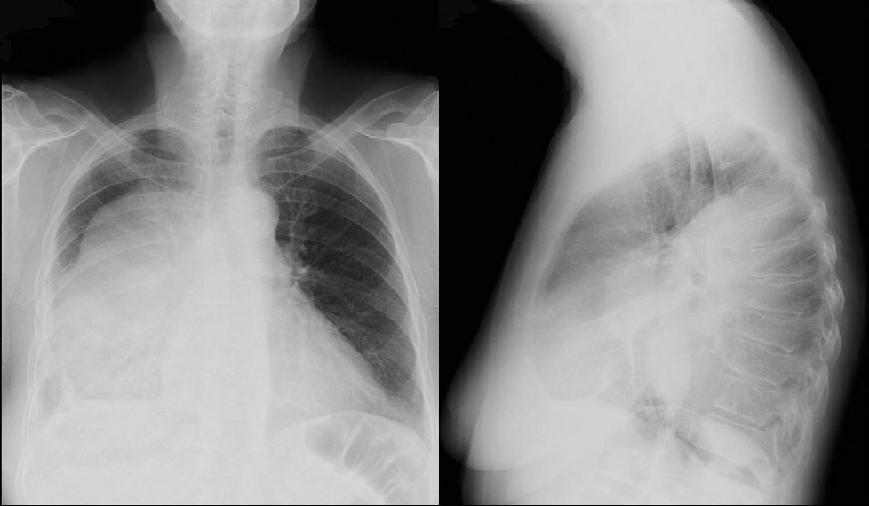
· Pleural-based opacity in non-dependent regions if loculation occurs.
· “D-sign” - loculated pleural fluid bulging out from the chest wall.
· Elliptical shape with well-delimited borders and conforms to the chest wall (Figs. 1; 2).
· Loculated collection of pleural fluid with or without gas pockets (Figs1; 2).
· Pleural air-fluid level may be seen when a bronchopleural fistula is present.
CT / MRI
· Stage I
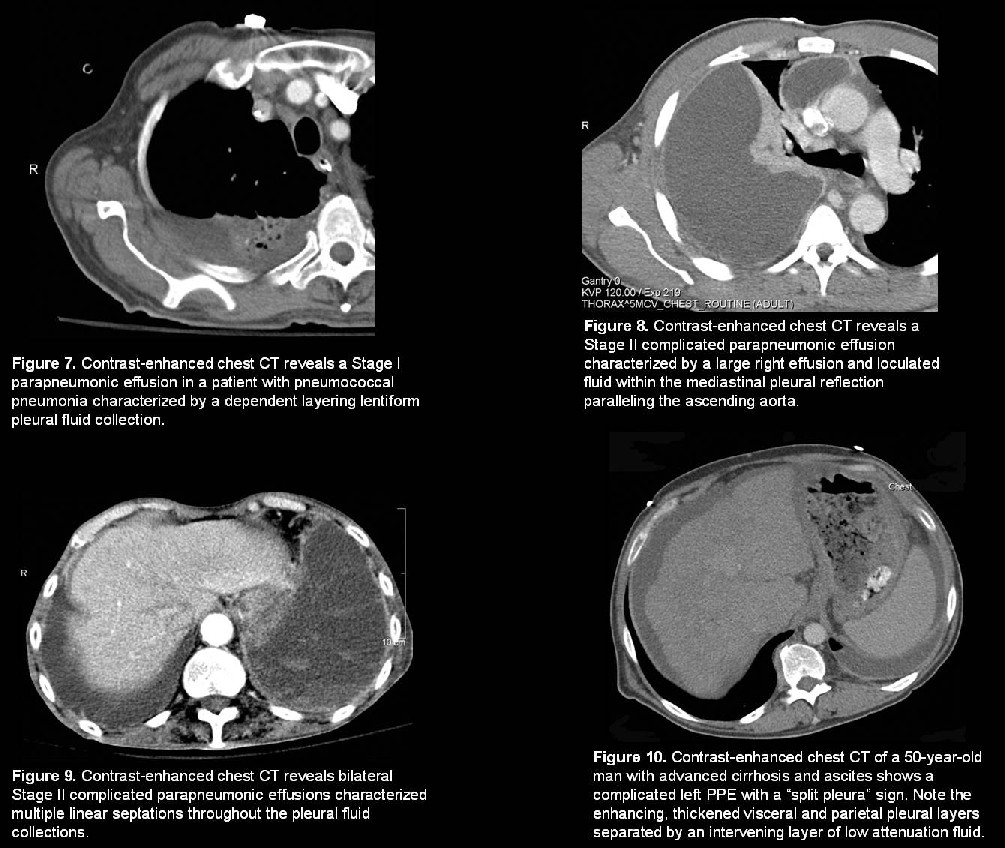
o Air-space disease associated with pleural effusion (Fig. 7).
o Lenticular fluid collection
· Stage II
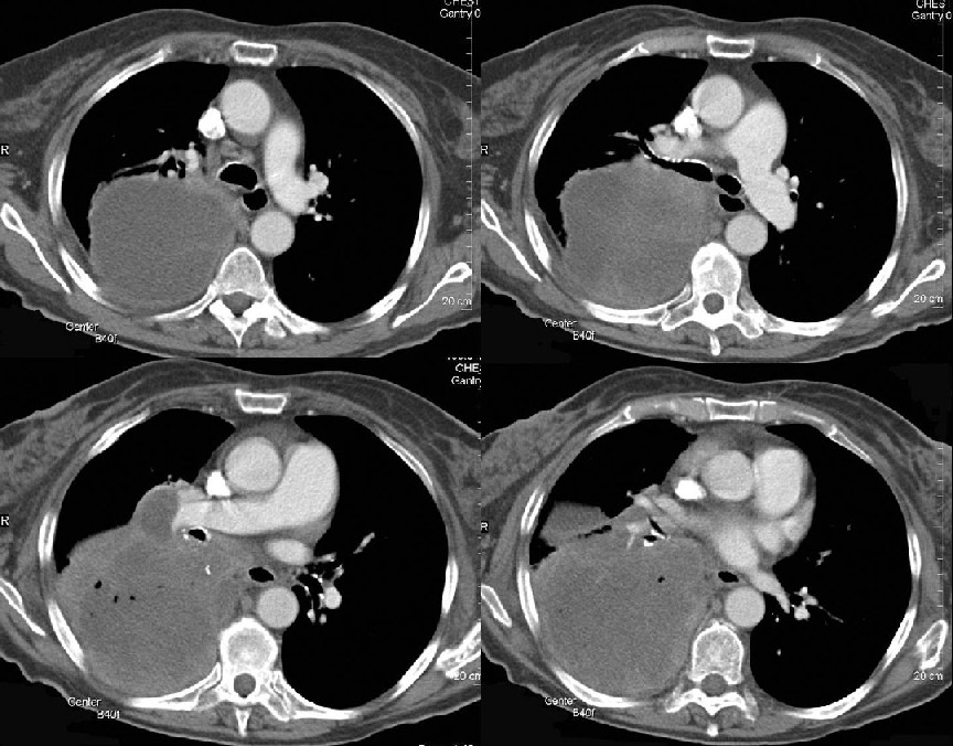
o Fixed, non-mobile pleural “mass” (Fig. 3-7).
o Pleural fluid in atypical locations (e.g., mediastinal pleural reflections; fissures) (Fig. 8).
o Thickening and or enhancement (i.e., hyperemia) of parietal pleura.
o Increased thickness and attenuation of costovertebral subpleural fat.
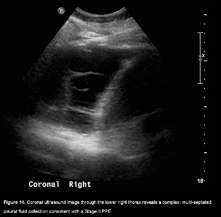
o Septations identified within the pleural fluid collection (Fig. 9).
o “ Split pleura sign” – enhancing, thickened visceral and parietal pleural layers separated by an intervening layer of low attenuation fluid (Fig. 10).
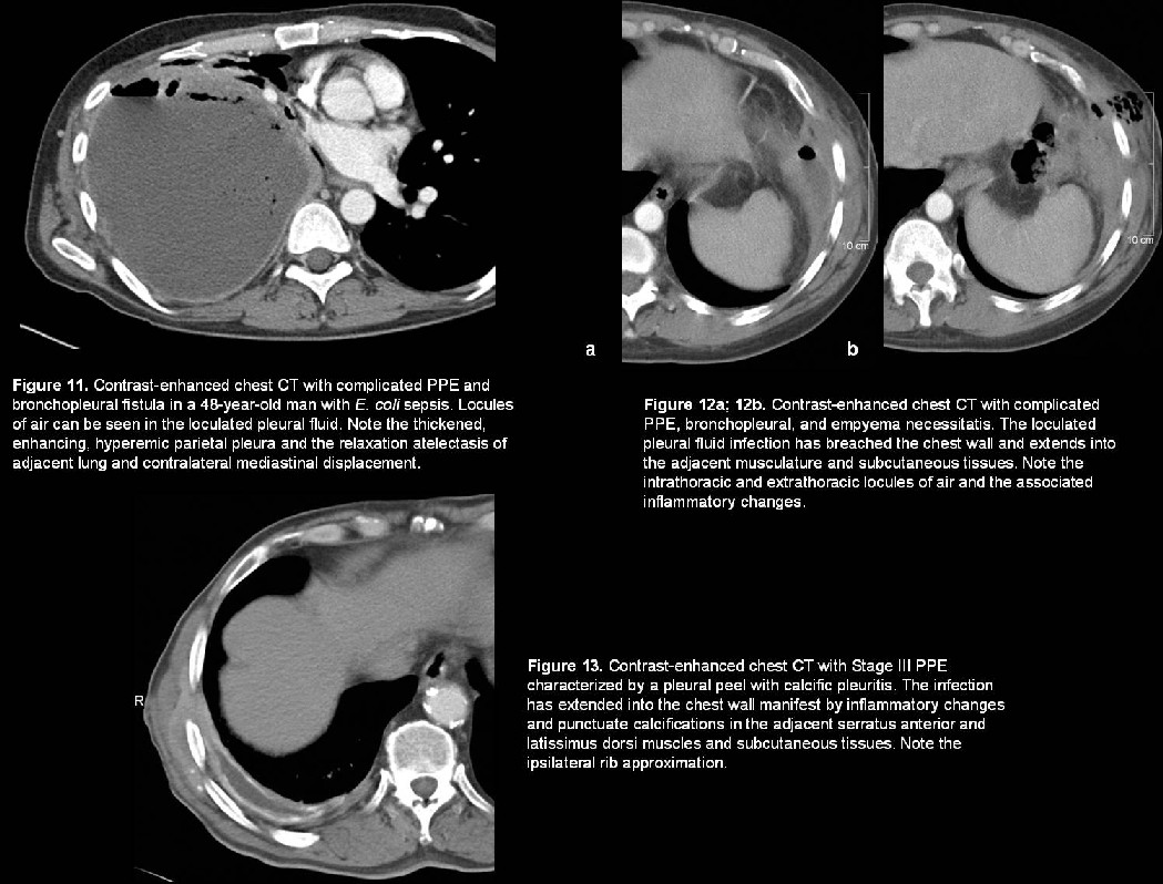
o Loculated gas bubbles in the pleural space with complicating bronchopleural fistula (Fig. 11).
o Empyema necessitatis when infected pleural fluid collection extends into chest wall (Fig. 12).
· Stage III
o Pleural rind or peel with or without calcific pleuritis (Fig. 13).
o Entrapment of the lung.
o Rib approximation and contraction of the affected hemithorax.
o Chest wall invasion or violation (Fig. 12; 13).
Ultrasound
· Important adjunct in defining the characteristics of the pleural fluid
· Detect small effusions
· Provide information about fluid viscosity
· Assess free-flowing versus loculation
· Detect the presence of septations (Fig. 14).
· Direct thoracentesis or catheter drainage
Treatment
The first priority is protecting the healthy lung. Empiric antibiotics are usually started before culture results have returned. The key to successful treatment is evacuation of the infected pleural space and elimination of the dead space in the pleural cavity. Treatment options include:
· Therapeutic thoracentesis
o Single thoracentesis and antibiotics may be effective in the earliest stage.
o If the fluid characteristics are borderline for chest tube placement, repeating a thoracentesis at 12-24 ° may be helpful.
o If the fluid LDH decreases and the pH and glucose increase, the patient is likely improving.
o If the LDH is increasing and the pH and glucose are decreasing, a chest tube should be placed.
· Tube thoracostomy
o Image-guided small bore caliber single or multiple 8-16F catheters for continuous drainage and or i ntrapleural instillation of fibrinolytics.
o Fibrinolytics often increase successful catheter drainage in fibropurulent or early organized cavities by decreasing fluid viscosity and lysing adhesions.
§ Streptokinase and urokinase are equally effective.
§ Streptokinase (250,000 U) in 30-60 ml normal saline given intrapleurally via chest tube. Clamp chest tube for 1-2 ° . Can be repeated for up to 14d.
§ Urokinase (100,000 U) diluted and administered in the same manner.
· Video assisted Thoracoscopic Surgery (VATS)
o Indicated for effusions with multiple loculations that are difficult to drain and those not responding to catheter drainage and stage III PPE.
o Evacuation of infected pleural fluid
o Lysis of adhesions
· Empyemectomy-Decortication and Open Drainage
o Indicated for late Stage II or Stage III PPE with inadequate pleural drainage after tube thoracostomy and intrapleural fibrinolytic therapy.
o Decortication preferred over open drainage
§ Full thoracotomy is performed.
§ All fibrous tissue is removed from the pleural surfaces.
§ All pus is evacuated from the pleural space.
o Open Drainage
§ Indicated for those patients too ill to tolerate decortication.
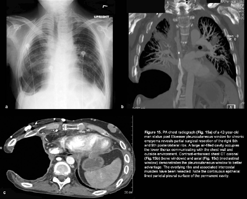
§ Segments of 1-3 ribs overlying the lower part of the empyema cavity are resected, and one or more short large-bore tubes are inserted into the cavity. The cavity is irrigated daily with antiseptic solution, and drainage from the tubes can be collected in a colostomy bag.
§ Pleurocutaneous window (Eloesser flap)
· Indicated for refractory pleural effusions (e.g., empyema, malignant effusions, esophagopleural fistulas).
· Creates permanent open access to the pleural space.
· Mid-axillary line horizontal incision; at least 2 ribs and the intervening intercostal muscles are removed; the skin is circumferentially sewn directly to the parietal pleura, creating a continuous epithelial surface and insuring dependent drainage without the need for tubes (Fig. 15).
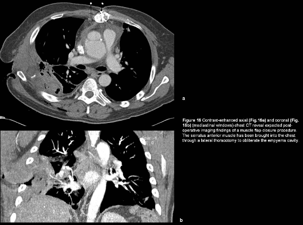
§ Muscle Flap Closure without Pleural Drainage
· Provides well-vascularized muscle tissue to close a bronchopleural fistula and obliterate the empyema cavity.
· Latissimus dorsi muscle
o Ideal bulk, pedicle length, and arc of rotation to fill most thoracic defects. May be used as a turn-over flap or advanced directly into the wound. Usually provides enough muscle bulk to obliterate an empyema cavity. Unfortunately, it has often already been divided in most antecedent open thoracic procedures.
· Serratus anterior muscle (Fig. 16).
o S econd most common muscle used to fill an empyema cavity. Muscle is thin enough to fill a small space and can be passed through a lateral thoracotomy incision. This muscle also can be brought into the chest with the latissimus dorsi muscle on a common pedicle.
· Pectoralis major muscle
o May be used as either a turn-over flap or placed directly in the wound. Intrathoracic placement requires creation of a window by partial rib resection of the 2nd or 3rd ribs to afford maximal length.
· Omentum
o May be used if the empyema space is not large or a well-vascularized reinforcement of a bronchopleural fistula is required. Advantages include its long reach, excellent vasculature, and its relative distance from the infectious process.
Prognosis
· Overall mortality is approximately 20%; advanced multilocular empyema can have a mortality of up to 50%.
· Empyema may be complicated by
o Necrosis of the visceral pleura; parietal pleural; and or chest wall.
o Bronchopleural or esophageal fistula.
o Osteomyelitis of the ribs or spine.
o Metastatic hematogenous systemic dissemination (e.g., brain abscess (es).
· Median time for healing treated by open-drainage procedures is 142 days.
PEARLS
· PPE is any pleural effusion associated with underlying pneumonia, lung abscess, or bronchiectasis.
· PPE is the most common cause of an exudative pleural effusion.
· Radiologic findings cannot identify a specific etiology; a specific diagnosis can only be made by isolation of the organism from the pleural fluid.
· Gram-negative anaerobic organisms are responsible for most culture-positive parapneumonic effusions in adults.
· Distinction between lung abscesses is important as empyema is treated with external drainage while lung abscess is often treated with antibiotics alone.
Suggested Readings
1. Arenas-Jiménez J, Alonso-Charterina S, Sánchez-Pavá J, Fernández-Latorre F, Gil- Sánchez S, Lloret-Llorens M. Evaluation of CT findings for diagnosis of pleural effusions. Eur Radiol 2000; 10(4):681-690.
2. Athanassiadi K, Gerazounis M, Kalantzi N. Treatment of post-pneumonic empyema thoracis. Thorac Cardiovasc Surg 2003; 51(6):338-341.
3. Huggins JT, Sahn SA, Heidecker J, Ravenel JG, Doelken P. Characteristics of trapped lung: pleural fluid analysis, manometry, and air-contrast chest CT. Chest 2007; 131(1):206-213.
4. Kearney SE, Davies CW, Davies RJ, Gleeson FV. Computed tomography and ultrasound in parapneumonic effusions and empyema. Clin Radiol 2000; 55(7):542-547.
5. Miller JI. The history of surgery of empyema, Thoracoplasty, Eloesser flap, and muscle flap transposition. Chest Surg Clin N Am 2000; 10(1):45-53, viii. Qureshi NR, Gleeson FV. Imaging of pleural disease. Clin Chest Med 2006; 27(2):193-213.
Filed under: Radiology, Medicine/Pulmonary
Original case written by its authors at Virginia Commonwealth University and published at this address as part of a weekly teaching collection. Reproduced here as an archive.