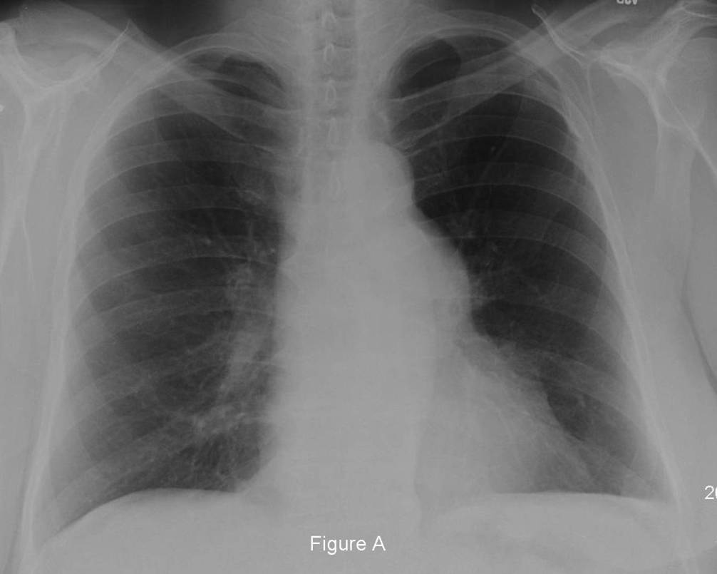
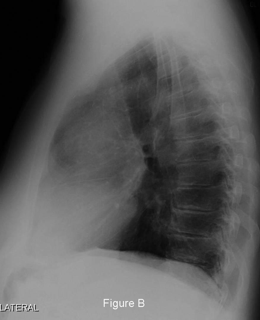
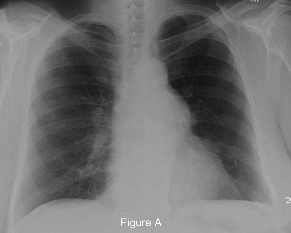
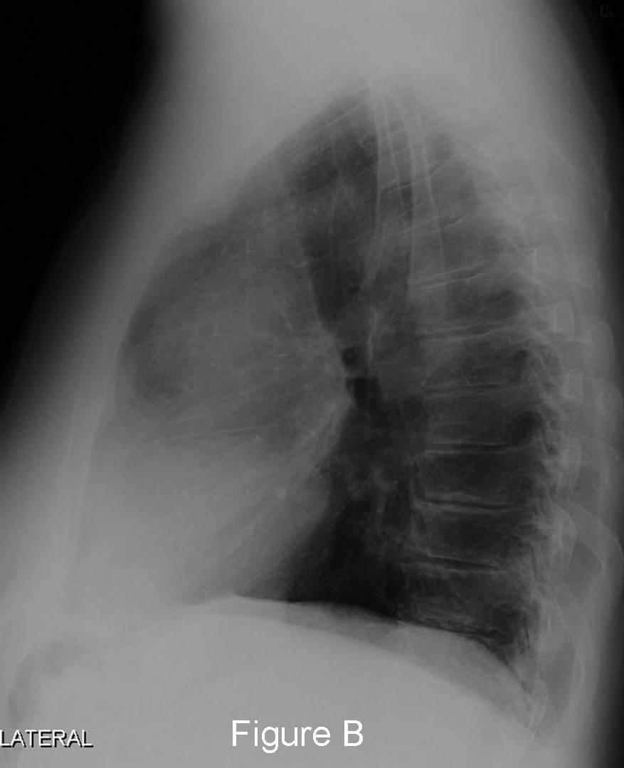
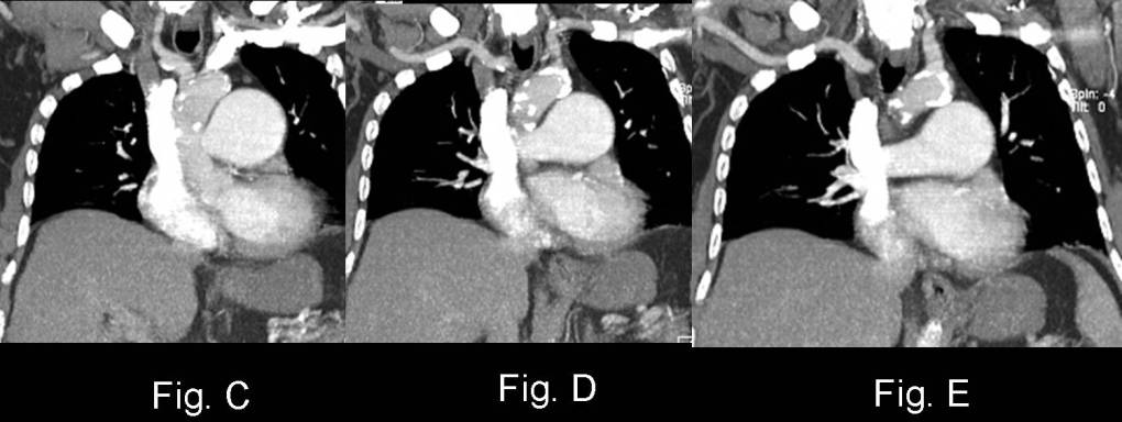
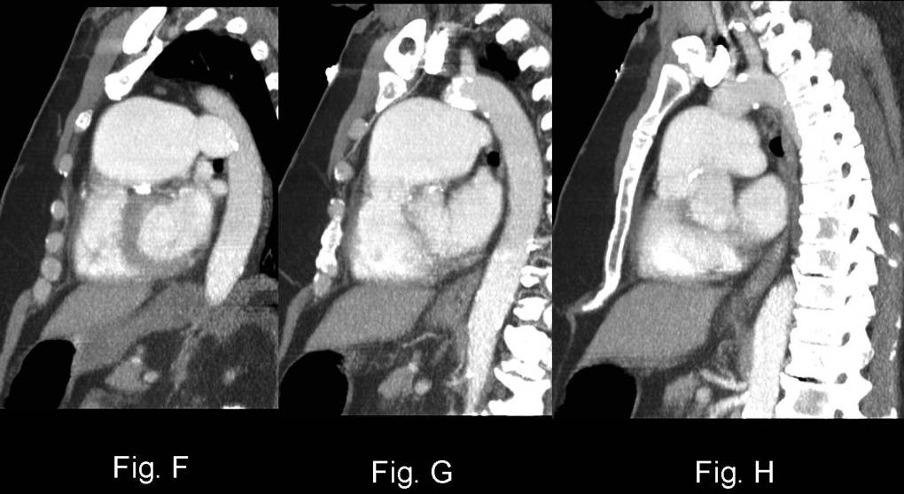
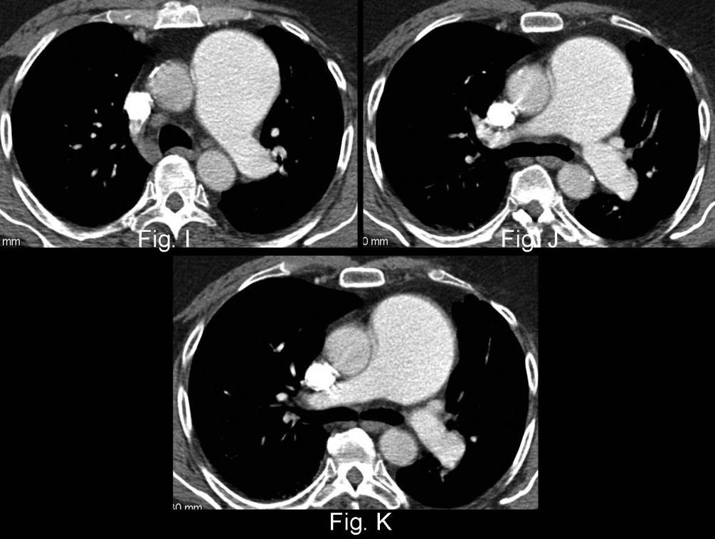
PA (Fig. A) and lateral (Fig. B) chest radiographs demonstrate a focal convex mass projecting over the left hilum that does not silhouette the left pulmonary artery. In fact, the left pulmonary artery can be seen at least a centimeter within the lateral edge of this opacity (“hilum overlay” sign) suggesting the mass is located in the anterior mediastinum. The lateral exam confirms the retrosternal – anterior mediastinal location of the apparent mass (Fig. B). On closer inspection, the frontal radiograph also shows that the pulmonary artery branches appear to converge toward the apparent hilar mass, suggesting the presence of an enlarged pulmonary artery (“hilum convergence” sign) (Fig. A). Contrast-enhanced CT coronal (PA radiograph equivalent) (Fig. C-E), sagittal (lateral radiograph equivalent) (Fig. F-H) and axial (Fig. I-K) images reveal the mass is formed by marked enlargement of the main pulmonary artery. Additionally, the left pulmonary artery is enlarged but the right pulmonary artery is normal is caliber. Diagnosis: Pulmonic Stenosis; “Hilum Overlay” and “Hilum Convergence” Sign
Differential Diagnosis
- Pulmonary artery hypertension
Background
Discussion
This case highlights the utility of the “hilum overlay” and the “hilum convergence” sign and the application of such in steering the differential diagnosis appropriately. The proximal segment of the left and right pulmonary arteries lie lateral to the cardiac silhouette or just within its outer edge in the vast majority of normal individuals. Felson described this normal apposition of the main pulmonary arteries to the lateral edge of the cardiopericardial silhouette as the rationale behind the “hilum overlay” sign. Specifically, the configuration of an anterior mediastinal mass can mimic an enlarged heart or pericardial sac. However, an anterior mediastinal mass cannot reside directly medial to the pulmonary artery because of the heart and pericardium. Therefore, a true mass will often overlap the main pulmonary arteries which can then be seen projecting within the margins of the mass. This sign should be contrasted with the “hilum convergence” sign. The latter aids in differentiating an enlarged pulmonary artery from a juxtahilar mediastinal mass. If the pulmonary artery branches converge toward the apparent mass, this suggests the presence of an enlarged pulmonary artery. On the other hand, if the pulmonary artery branches converge toward the heart, then a juxtahilar mass is likely present.
Etiology
Pulmonic stenosis is the most common congenital anomaly that produces obstruction of the right ventricular outflow tract and results from congenital malformation of the pulmonic valve. It occurs as an isolated anomaly in 7% of patients with congenital heart disease. It is also a common congenital cardiac lesion manifesting in adulthood.
Clinical Findings
Most patients are acyanotic and many are completely asymptomatic. Symptomatic patients most often present in the 3rd-4th decade of life. On auscultation, a harsh long systolic ejection murmur may be heard.
Imaging Findings
Conventional Radiography
- Mild-moderate enlargement main pulmonary artery
- Left pulmonary artery enlargement
- Normal size right pulmonary artery, peripheral pulmonary arteries and pulmonary veins
- Normal size heart
- Right ventricular failure may be associated with right ventricular enlargement
- Pulmonic valve calcification (rare)
MDCT / Cardiac MRI
- Main and left pulmonary artery enlargement
- Normal size right pulmonary artery
- Turbulent blood flow in GRE sequences
- Differential blood flow in the pulmonary arteries across the stenotic valve (VENC sequences)
Management
- Balloon dilation
- Valve replacement
Prognosis
- Good with appropriate treatment
- Increased morbidity and morality from infective endocarditis
Caveats
Pulmonic stenosis (PS) and pulmonary artery hypertension (PAH) may both manifest with pulmonary artery enlargement. However, PS will be characterized by enlargement of the main and left pulmonary artery and a normal sized right pulmonary.
Selected Readings
- Felson BF. Localization of Intrathoracic Lesions In: Chest Roentgenology 1973; W.B. Saunders, Philadelphia: 39.
- Parker MS, Rosado-de-Christenson ML, Abbott GF. Pulmonic Stenosis In: Teaching Atlas of Chest Imaging 2006; Thieme, New York: 96-99.
Original case written by its authors at Virginia Commonwealth University and published at this address as part of a weekly teaching collection. Reproduced here as an archive.