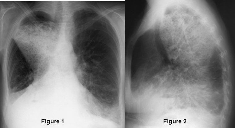
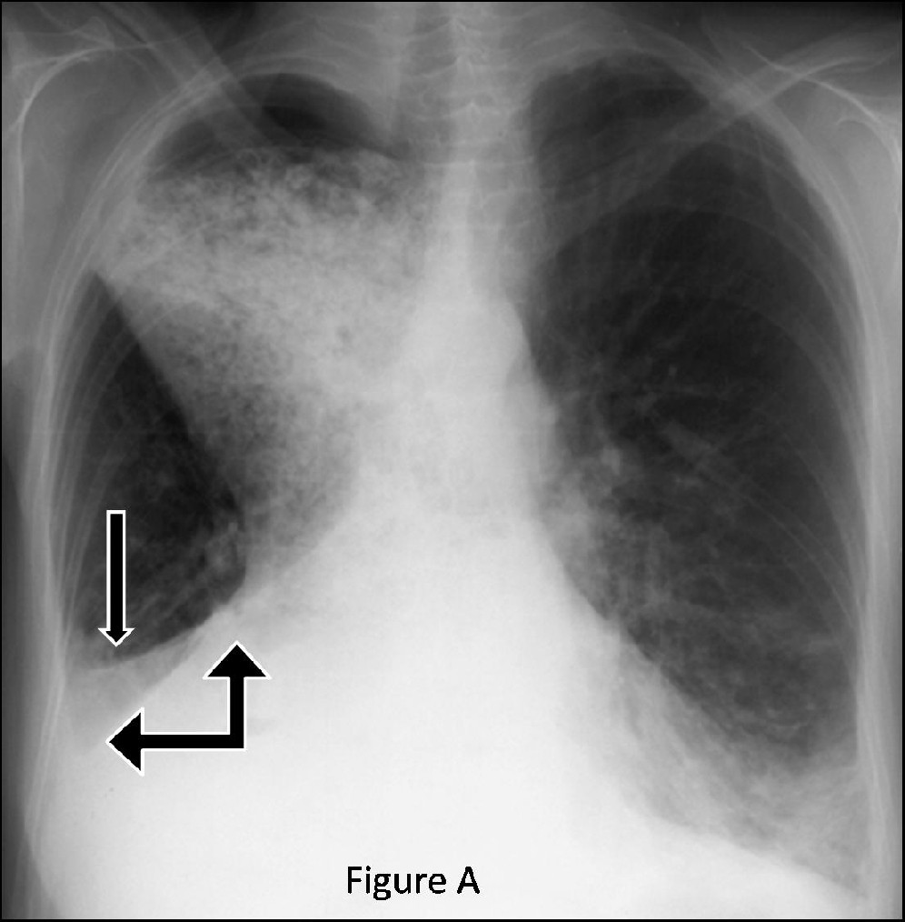
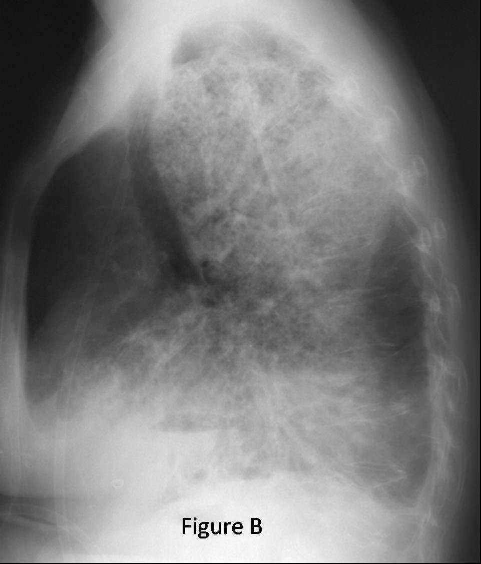
Clinical Presentation: Elderly woman with dysphagia and halitosis with diminished breath sounds over the right chest (originally withheld). Radiologic Findings: PA (Fig. A) and lateral (Fig. B) chest radiographs demonstrate two primary radiographic abnormalities of concern. The first is that of combined right middle lobe and right lower lobe atelectasis. The right hemithorax is relatively smaller in volume compared to the left from the underlying volume loss and there is ipsilateral mediastinal shift. Note the downward displacement of the horizontal fissure (single arrow) and right heart border silhouette from the middle lobe collapse. Notice also the downward displacement of the oblique fissure (double head right angle arrow), the small and inapparent right hilum and interlobar descending pulmonary artery, and silhouette of the diaphragm; cardinal signs of right lower lobe collapse. The second pertinent radiologic finding is a mottled, mixed density, mediastinal opacity, eccentric to the right, which extends from the T3 level to the hiatus. This mass displaces the trachea anteriorly and obliterates the retrotracheal triangle, retrocardiac clear space, and inferior hilar window consistent with a massively distended, fluid and debris-filled esophagus. Diagnosis: Combined Right Middle and Lower Lobe Atelectasis; Esophageal Achalasia
Differential Diagnosis
- Mucus plug or other obstructing foreign body of the bronchus intermedius
- Primary and secondary endobronchial lesions obstructing the bronchus intermedius
- Other causes of extrinsic compression of bronchus intermedius
- Other mediastinal based lesions
- Lymphadenopathy
- Other diseases of the esophagus resulting in obstruction
- Distal neoplasia
- Strictures
- Chagas disease
Discussion
Collapse of multiple lobes reflects the sum of the signs of collapse of the individual lobes. Combined right middle lobe (RML) and right lower lobe (RLL) collapse or atelectasis can be explained by a single intrinsic lesion obstructing or extrinsic lesion compressing the single conduit to both these lobes, the bronchus intermedius. In this case, the bronchus intermedius is extrinsically compressed by the massively dilated and distended fluid and debris filled esophagus resulting from achalasia causing the combined multi-lobar volume loss.
Imaging Findings (Figure A and Figure B)
Sum of findings associated with collapse of the individual lobes
Isolated RML Collapse:
Frontal Radiography
- Silhouette of right heart border
- Minor fissure displaced inferiorly
Lateral Radiography
- Well-defined, curvilinear opacity bordered by oblique and horizontal fissures, extending anteriorly and inferiorly from the hilum
- Collapsed middle lobe may be very thin and misinterpreted as a thickened fissure
Isolated RLL Collapse:
Frontal Radiography
- Partial or complete silhouette of right hemidiaphragm, paraspinal interface, and inferior cava depending on degree of volume loss; right heart border remains visible
- Oblique fissure displaced inferiorly and medially but maintains a convex lateral border
- Small or in apparent ipsilateral hilum and interlobar pulmonary artery
- Inferior displacement of the main stem bronchus
- Reorientation of main stem and lower lobe bronchi into a more vertical plane
- Narrowing of the carinal angle
- Opaque lobe assumes a triangular morphology
- Apex directed toward hilum
- Base directed toward hemidiaphragm
Lateral Radiography
- Upper half of the oblique fissure shifts inferiorly and lower half posteriorly
- Horizontal fissure may or may not shift
- Atelectatic lobes create an opacity over the inferior hemithorax extending from anterior to posterior chest wall
Caveat
- Combined RML and RLL collapse may be misinterpreted as isolated RLL collapse
- If the opacity extends to the costophrenic angle, combined collapse is likely
- If the horizontal fissure is visualized in its normal anatomic position, isolated RLL collapse is present
- Combined RML and RLL collapse may be misinterpreted as a right pleural effusion
- Identification of both the horizontal and oblique fissures in their normal anatomic positions favors effusion
- Preservation of the lateral costophrenic sulcus argues against pleural effusion
Selected Readings
- Parker MS, Rosado-de-Christenson ML, Abbott GF. Right Middle Lobe Atelectasis. In: Teaching Atlas of Chest Imaging. New York, Thieme; 2006: 188-190.
- Parker MS, Rosado-de-Christenson ML, Abbott GF. Right Lower Lobe Atelectasis. In: Teaching Atlas of Chest Imaging. New York, Thieme; 2006: 196-198.
- Parker MS, Rosado-de-Christenson ML, Abbott GF. Combined Right Middle and Right Lower Lobe Atelectasis. In: Teaching Atlas of Chest Imaging. New York, Thieme; 2006: 199-200.
Original case written by its authors at Virginia Commonwealth University and published at this address as part of a weekly teaching collection. Reproduced here as an archive.