Archived case 38 · Mar 11-Mar 18, 2010
Constrictive Pericarditis
18-year-old man with dyspnea
The question posed to readers
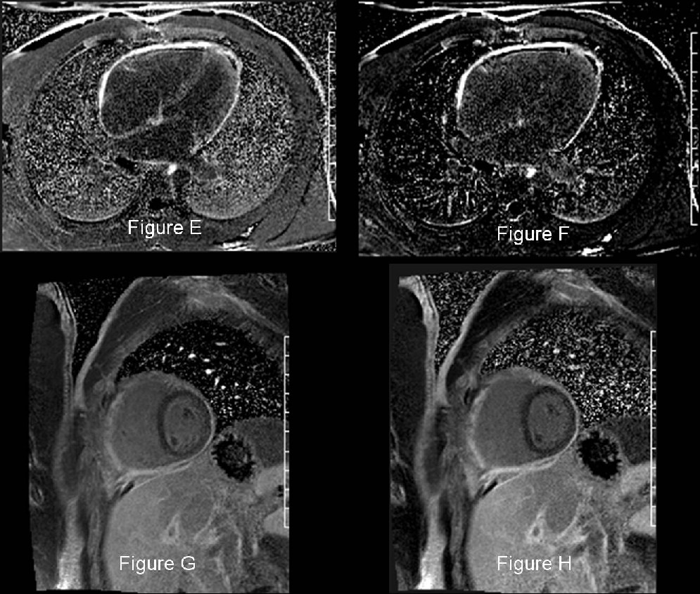
What are the pertinent imaging findings? What is your diagnosis? What are some possible clinical causes for this finding?
Archived case 38 · Mar 11-Mar 18, 2010
18-year-old man with dyspnea
What are the pertinent imaging findings? What is your diagnosis? What are some possible clinical causes for this finding?
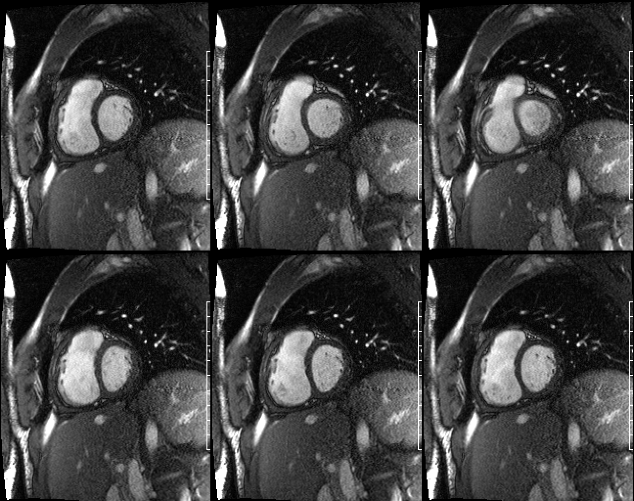
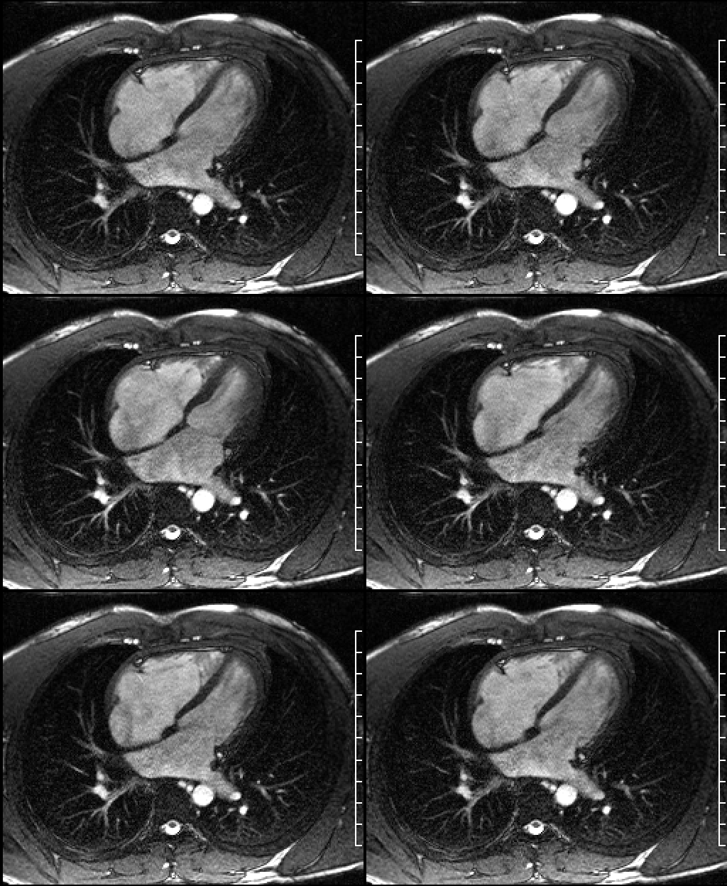
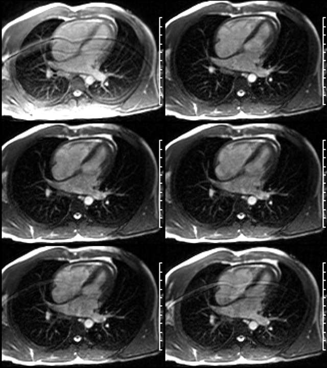
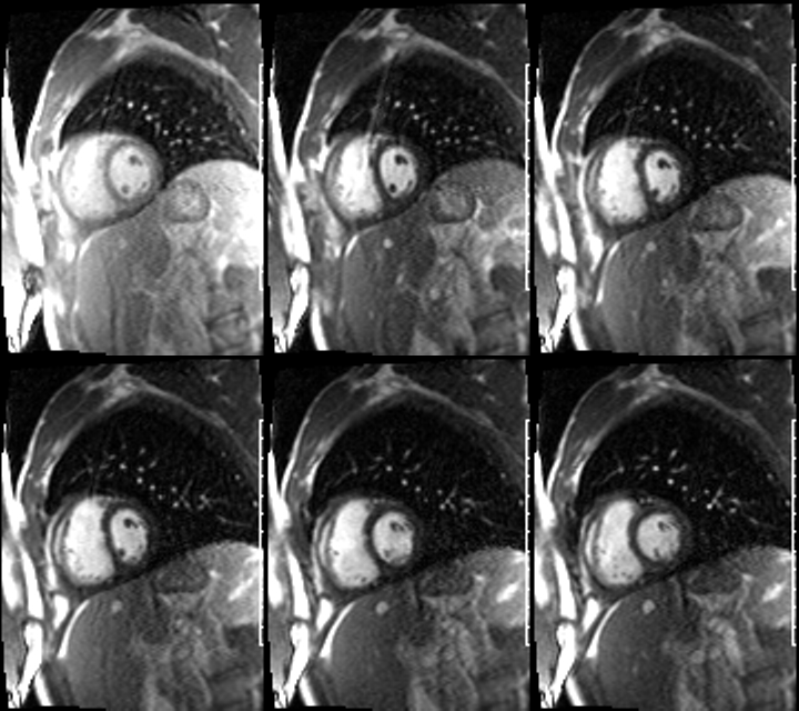
Constrictive Pericarditis
Differential Diagnosis
None
Background
Discussion
The normal pericardium is composed of 2 layers: a tough fibrous parietal pericardial layer and a smooth visceral pericardial layer. Approximately 50 mL of transudative fluid is normally present between these 2 layers (i.e., intrapericardial space) which minimizes friction during the cardiac cycle. Inflammation of the pericardial layers is referred to as pericarditis. Acute and subacute pericarditis may be associated with fibrin deposition along these pericardial layers and a concomitant pericardial effusion. Subsequent organization results in fibrosis, scarring, and sometimes calcification, most often of the parietal pericardium. The thickened, fibrotic pericardium then impairs normal late diastolic filling of the ventricle. Approximately 9% of patients with acute pericarditis go on to develop constrictive physiology such as demonstrated in this case.
Etiology
Those clinical entities associated with acute pericarditis are likewise responsible for the development of constrictive pericarditis. These most commonly include: idiopathic (presumably viral) (e.g., coxsackievirus A and B, adenoviruses); tuberculosis; following cardiac surgery; and radiation-therapy induced. Less common causes include: neoplasia; uremia; various connective tissue disorders (e.g., rheumatoid arthritis, systemic lupus erythematosus, scleroderma); drug-induced (e.g., Procainamide, Hydralazine); and following myocardial infarction.
Clinical Findings
Dyspnea is the most common presenting symptom in virtually all patients. Fatigue and orthopnea are not uncommon. Patients may also experience lower extremity edema. Unexplained jugular venous distention, pleural effusion, hepatomegaly, and or ascites may be evident on physical exam. Kussmaul sign (ie, elevation of systemic venous pressures with inspiration) is a common but nonspecific finding.
Imaging Findings
Conventional Radiography
MDCT
MRI
Treatment
Surgical pericardiectomy is the procedure of choice in the appropriate patient. However, in cases where constriction has been present for a prolonged period of time, the response may be less dramatic, due to the development of extensive atrophy and fibrosis.
Prognosis
Symptoms following pericardiectomy commonly improve, but evidence of abnormal diastolic filling often remains. Only 60% of patients have complete normalization of cardiac hemodynamic function.
Caveats
Selected Readings
Filed under: Radiology, Medicine/Pulmonary
Original case written by its authors at Virginia Commonwealth University and published at this address as part of a weekly teaching collection. Reproduced here as an archive.