Diagnosis: Swyer-James (MacLeod) syndrome
Differential Diagnosis Unilateral Hyperlucent Thorax
Technical
- Rotation
- Lateral decentering
Altered Chest Wall Anatomy
- Mastectomy
- Rectus abdominus muscle flap transposition (TRAM)
- Pectoralis muscle flap transposition
- Congenital absence pectoralis muscle (Poland syndrome)
- Scoliosis
Vasculature
- Acute pulmonary thromboembolic disease
- Pulmonary artery hypoplasia / stenosis /absence
Pleural Disease
Large and Small Airways
- Emphysema
- Large bulla / pneumatocele
- Central obstructing tumors / endobronchial lesions
- Aspirated foreign body
- Post-infectious bronchiolitis obliterans (Swyer-James / MacLeod syndrome)
- Congenital lobar hyperexpansion (emphysema) of the lung
- Following pneumonectomy / unilateral lung transplant
Background
Discussion
Swyer-James or MacLeod syndrome is a post-infectious constrictive bronchiolitis that is usually the sequelae of severe childhood pneumonia. Adenovirus types 3, 7, and 21 have been implicated most commonly, but other infectious etiologies include: Mycoplasma pneumoniae, parainfluenza virus types 1-3, influenza virus types A and B, respiratory syncytial virus, measles, and Bordetella pertusis.
Etiology
It is postulated that the infectious acute bronchiolitis progresses to fibrous obliteration of the airway lumen, namely damaging the terminal and respiratory bronchioles, preventing normal development of their alveolar buds. The affected lung tissue therefore becomes “stunted” and “underdeveloped”, both in terms of volume and blood flow and circulation. The peripheral lung parenchyma is usually unaffected and remains inflated via collateral pathways (e.g., incomplete fissures, pores of Kohn, canals of Lambert) and air trapping ensues. This process may involve an entire lung, a lobe or lobes or just a segment.
Clinical Findings
Most adult patients are asymptomatic. Symptomatic patients may experience chronic cough, repeated pulmonary infections, dyspnea, decreased exercise tolerance, fatigue, and hemoptysis.
Imaging Findings
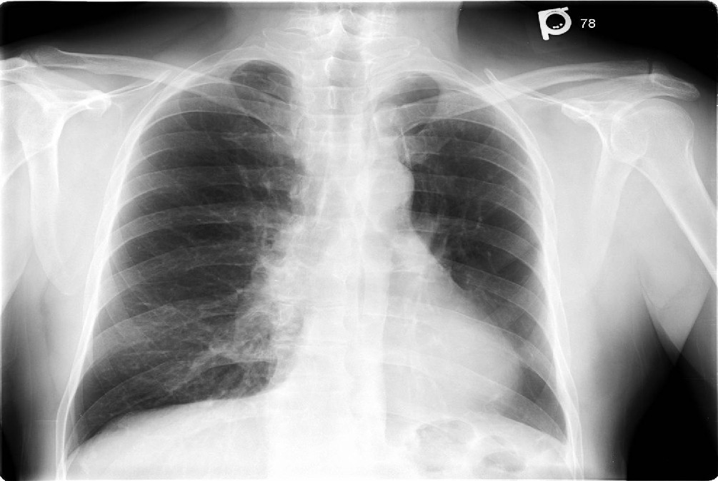
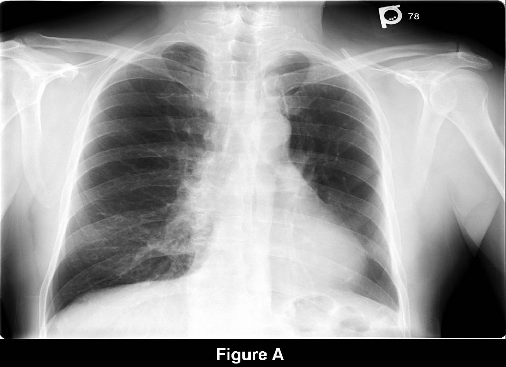
Conventional Radiography (Fig. A)
- Unilateral hyperlucent lung
- Small or normal-sized hemithorax
- Overinflation of contralateral lung
- Diminutive pulmonary hilum
- Decreased peripheral pulmonary arteries
- Mediastinal shift toward the hyperlucent thorax
Ventilation-Perfusion (V/Q) Scan
- Decreased perfusion on affected side
- Retention of inhaled radionuclide on affected side
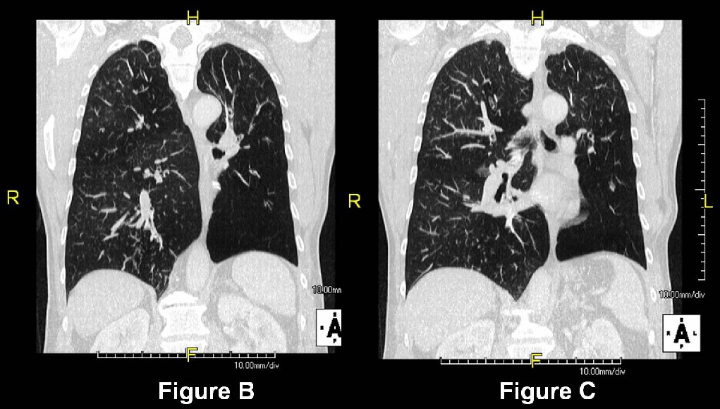
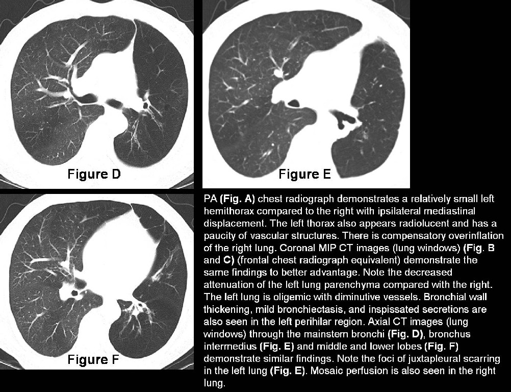
MDCT (Fig. B-F)
- Small or normal sized lung with patent bronchial tree
- Diminished or normal lung attenuation without the normal anteroposterior attenuation gradient
- Small central and peripheral pulmonary arteries
- Bronchiectasis
- Inspissated secretions
- Juxtapleural scarring and or atelectasis
- Air-trapping on expiratory images
- Mosaic perfusion patterns
Caveats
On chest radiography, this syndrome must be differentiated from an endobronchial lesion incompletely obstructing the lumen of a main or lobar bronchus, unilateral bullous emphysema, and various pulmonary artery abnormalities. CT is the optimal tool to make this differentiation.
Selected Readings
- Marti-Bonmati L, Perales FR, Catala F, Mata JM, Calonge E. CT findings in Swyer-James syndrome. Radiology 1989; 172(2): 477-480.
- Müller NL. Unilateral hyperlucent lung: MacLeod versus Swyer-James. Clin Radiol. 2004; 59(11):1048.
- Travis WD, Colby TV, Koss MN, Rosado-de-Christenson ML, Müller NL, King TE Jr. Bronchiolar Disorders. In: King DW, ed. Atlas of Nontumor Pathology: Non-Neoplastic Disorders of the Lower Respiratory Tract, first series, fascicle 2. Washington , DC : American Registry of Pathology; 2002: 369.
Original case written by its authors at Virginia Commonwealth University and published at this address as part of a weekly teaching collection. Reproduced here as an archive.