Thoracic Imaging Archive

Archived case 3 · Jun 10-Jun 17, 2009

Acute Pulmonary Embolism with Peripheral Infarct

90 year old patient with right upper quadrant pain

The question posed to readers

Please provide findings and differential diagnosis
Images

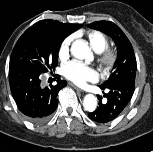
Radiograph 1 from archived case 3
Figure 1
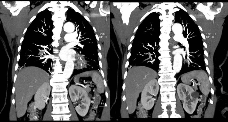
Radiograph 2 from archived case 3
Figure 2
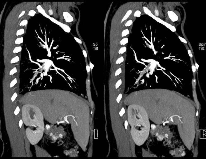
Radiograph 3 from archived case 3
Figure 3
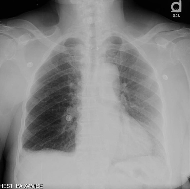
Radiograph 4 from archived case 3
Figure 4

Diagnosis

Acute Pulmonary Embolism with Peripheral Infarct

Hyperlucent right lung with perpherial opacity in the right lung base. Westermark Sign and Hampton's Hump - see below for descriptions of these signs.     Diagnosis: Acute Pulmonary Embolism with Peripheral Infarct

Differential Diagnosis – Unilateral Hyperlucent Lung

  Increased Transradiancy of Chest Wall Origin (Spurious)

  • Congenital absence of pectoralis major muscle
  • Mastectomy
Congenital Anomalies
  • Absence or hypoplasia of one pulmonary artery
  • Pulmonary agenesis
  • Extrapulmonary sequestration
Intrinsic Obstruction of One Pulmonary Artery
  • Embolus
  • Tumor
Extrinsic Compression of One Pulmonary Artery
  • Tumor
  • Nodes
Compensatory Hyperinflation
  • Atelectasis Status after lobectomy
Partially Obstructed Airways Intrinsic Obstruction of Major Bronchus
  • Tumor—benign or malignant
  • Postinflammatory stricture
  • Foreign body
  • Amyloid
Extrinsic Obstruction
  • Congenital   
    • Aberrant vessel   
    • Extrapulmonary sequestration
  • Tumor
  • Nodes
Congenital Malformation of Bronchus Causing "Congenital Emphysema" Unilateral Acquired Bronchitis and Bronchiolitis Obliterans
  • Swyer-James-Macleod syndrome

Parenchymal Findings in Acute PE

Signs on Chest Xray suggestive of PE

Westermark Sign: Oligemia (vasoconstriction) seen distal to a pulmonary embolus.
Hampton's hump: A wedge shaped, pleural based consolidation associated with pulmonary infarction.
Fleischner sign: Prominent central pulmonary artery

Statistically significant findings associated with PA

  • wedge shaped parenchymal consolidation
  • linear bands

No statistical difference in prevalence of non wedge shaped consolidation, areas of decreased attenuation, atelectasis, or pleural effusion

Coche et al. Radiology 1998

Parenchymal Consolidation in PE

Hemorrhage:

  • Ischemic damage to endothelial and alveolar epithelial cells
  • Red blood cells and fluid enter into air spaces

Hemorrhage with Infarct:

  • Occurs in 10-15% of PE
  • Pleural based opacity with apex directed toward hilum
  • May contain central areas of low attenuation representing uninfarcted secondary pulmonary lobules

Pneumonia

  • Seen with septic emboli or superimposed infection of infarcted lung

Hemorrhage vs. Infarct

  • Consolidation rapidly clears (several days) in hemorrhage
  • Consolidation slowly clears (weeks) in infarct which may give rise to linear bands

References

  • E.E. Coche, N.L. Muller, K.I. Kim, B.R. Wiggs, and J.R. Mayo 
    Acute pulmonary embolism: ancillary findings at spiral CT 
    Radiology 1998 207: 753-758.
  • Robert G. Fraser , J. A. Peter Par, Diagnosis of diseases of the chest, Saunders 1977
  • http://www.mdconsult.com/das/book/body/143494216-2/0/1288/321.html

Filed under: Radiology, Medicine/Pulmonary

Original case written by its authors at Virginia Commonwealth University and published at this address as part of a weekly teaching collection. Reproduced here as an archive.

Back to the case index