Diagnosis: Remote thoracoplasty and partially calcified oleothorax for tuberculosis
Differential Diagnosis
- Loculated pleural effusion
- Organized empyema
- Hemothorax with calcification
- Pleural neoplasia (e.g., mesothelioma)
Discussion
Background
In the pre-antibiotic era, lung and pleural infection by the obligate aerobe mycobacterium tuberculosis was treated with various forms of collapse therapy. The various forms of collapse therapy included: the induction of an artificial apical pneumothorax with resultant upper lobe collapse; thoracoplasty; and plombage. Pneumothorax was the first effective form of collapse therapy and attained world-wide acceptance as the standard and almost universally preferred method of pulmonary collapse. Surgical thoracoplasty was described by John Alexander in the 1930s. This surgical procedure entailed the sequential subperiosteal resection of the posterolateral aspect of numerous ribs and transverse processes. The initial surgery involved resection of the1st-3rd ribs. Subsequent surgeries might necessitate resecting the 4th through as much as the 7th ribs. Without the chest wall skeletal support, the apical and upper lobe lung parenchyma would collapse against the mediastinum, eliminating effective gas-exchange, essentially starving the obligate aerobes for oxygen. Success was achieved in 75-93% cases; however the operative mortality was as high as 10%. Plombage involved the extrapleural insertion of a “plombe” to collapse the lung. Employed plombes included fat; oils; solid paraffin wax; Lucite spheres; plastic ping pong balls; and sponges of inert plastic material. Oleothorax is the instillation of vegetable or mineral oil into the pleural or extrapleural space. It was often used as an adjunct to collapse therapy for the treatment of tuberculosis and tuberculous empyema during the second quarter of the 20th century in the United States. This procedure was largely abandoned in the early 1950’s with the advent of effective pharmacotherapy.
Clinical Findings
Collapse therapy was effective in controlling not only patient symptoms such as cough and hemoptysis, but also the infection as well, rendering positive sputum cultures negative in a high percentage of cases. However, the resultant chest wall deformity was undesirable in many, especially younger patients, as was the sequelae of restrictive physiology and dyspnea that often ensued. The instilled oil in oleothorax was usually left in place for 18-24 months, allowing the infected lung to “rest” and “recover”. Over half of the patients undergoing oleothorax developed an intense pleuritis associated with fever and prostration that necessitated aspiration of the oil. However, for the treatment of tuberculous empyema, this intense pleural reaction was viewed as a positive clinical sign indicating “cleansing” of the pleural space. Many such patients did improve clinically, were subsequently lost to follow-up, and never had the oil removed. These latter patients are occasionally incidentally encountered on imaging studies performed today.
Imaging Findings
Conventional Radiography
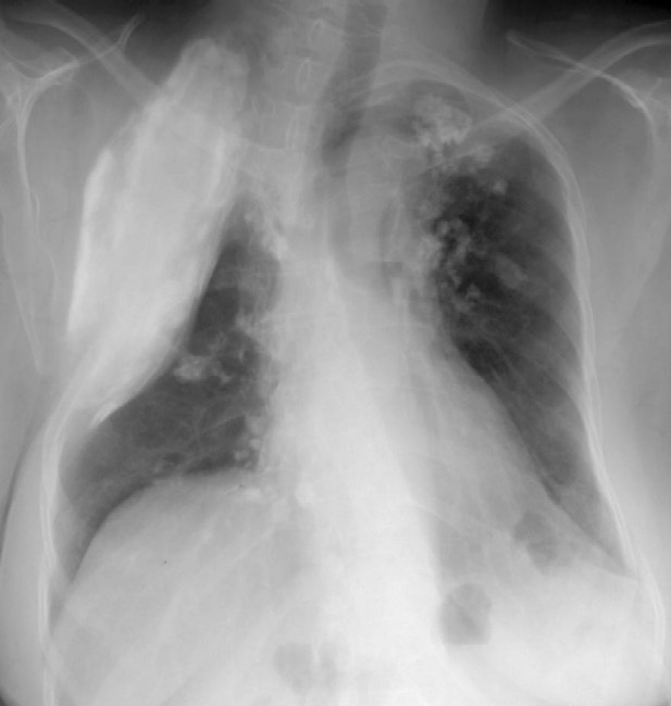
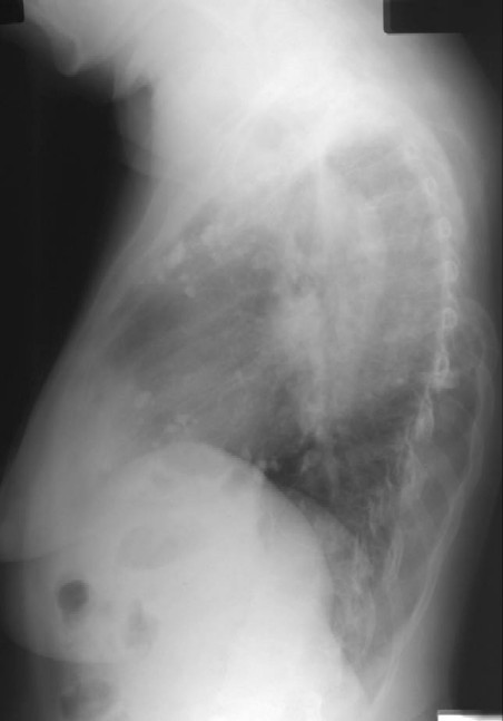
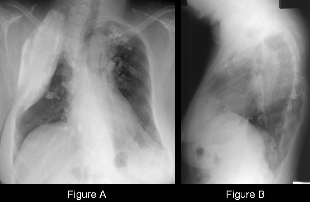
- Pathognomonic deformity of the upper thoracic cage (Fig. A and Fig. B)
- More cephalad ribs appear truncated or absent; corresponding transverse processes may also be absent (Fig. A and Fig. B)
- Remaining ribs may appear distorted and vertically oriented
- Volume of affected hemithorax is significantly diminished; upper lobe is airless (Fig. A and Fig. B)
- Mediastinal shift occurs towards the side of the thoracoplasty (Fig. A and Fig. B)
- High density extrapulmonary intrapleural or extrapleural lesion +/- calcification (Fig. A and Fig. B)
- Sequelae of the previous granulomatous insult manifest by pleural and parenchymal fibrosis; calcified lung nodules and lymph nodes (Fig. A and Fig. B)
CT
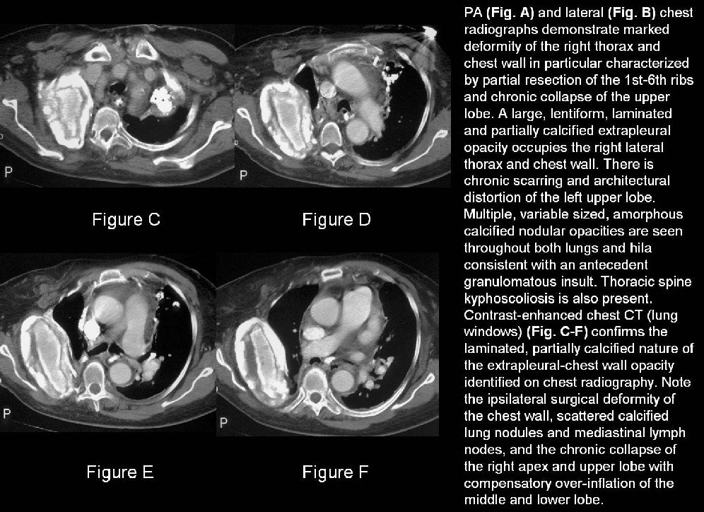
- Many of the same imaging features seen on conventional radiography (Fig. C-F)
- Laminated; partially calcified intra- or extrapleural space occupying mass (Fig. C-F)
- Regions of irregular pleural thickening and or calcification sequelae of the pleuritis
- Triphasic encased layering intra- or extrapleural fluid collection may also be seen with oleothorax
- Radiolucent buoyant layer that floats anteriorly (-162HU) (pure lipid)
- Intermediate layer of oil emulsion and serous fluid (-43HU)
- Dependent layer of aqueous-cellular suspension (36HU)
Present day Indications for Thoracoplasty
- Tuberculous empyema
- Drug resistant tuberculosis; in the setting of ineffective or impossible lung resection
- Refractory pyogenic empyema
- Post-operative empyema with bronchopleural fistula
- Infected post-resection space not closing with drainage alone, and concomitant tailoring thoracoplasty in conjunction with lung resection
Complications associated with Oleothorax
- Pleural calcification (Fig. A-F)
- Bacterial empyema
- Bronchopleural fistula
- Pleurocutaneous fistula
- Expansion
- Acute-respiratory distress
- Chronic-asymptomatic
Caveats:
- The pleural or extrapleural oleothorax may “expand” without either reactivation tuberculosis or development of empyema and may occur years after the instillation.
- No air should be present within the affected pleural or extrapleural collection; the presence of such should alert the physician to the possibility of underlying empyema or bronchopleural fistula.
- With the emergence of multi-drug resistant tuberculosis, thoracoplasty may someday again be required, with greater frequency, in the primary treatment of tuberculosis.
Selected Readings
- Deboisblanc BP, Burch WC, Buechner HA, Haponik EF. Thorax 1988; 43: 572-573.
- Fahy RJ, Morales J, King M. Late reactivation of tuberculosis in an oleothorax. J Thorac Imaging 2004; 19(1):35-37.
- Hutton L. Oleothorax: expanding pleural lesion. AJR 1984; 142: 1107-1110.
- Kumar H. Artificial pneumothorax in the treatment of pulmonary tuberculosis. Chest 1958; 33: 335-336.
- Meyer WL. Oleothorax. Chest 1947; 13: 467-470.
Original case written by its authors at Virginia Commonwealth University and published at this address as part of a weekly teaching collection. Reproduced here as an archive.