Usual Interstitial Pneumonia (UIP); (idiopathic pulmonary fibrosis) (IPF) Aka cryptogenic fibrosing alveolitis
Diagnosis: Usual Interstitial Pneumonia (UIP); (idiopathic pulmonary fibrosis) (IPF) Aka cryptogenic fibrosing alveolitis
Differential Diagnosis
- Mixed connective tissue and collagen vascular disorders with IPF
- Recurrent bouts of aspiration
- Chronic pulmonary drug toxicity
- Asbestosis
- Chronic hypersensitivity pneumonitis
- Fibrosing NSIP
Discussion
Background
Usual interstitial pneumonia (UIP) is the most common of the idiopathic interstitial pneumonias (IIPs). It is a histologic pattern of chronic fibrosing interstitial pneumonia that may be idiopathic (idiopathic interstitial pneumonia, IPF) or may be a manifestation of various connective tissue disorders (e.g., rheumatoid arthritis, scleroderma), cytotoxic (e.g., Bleomycin, Busulfan, Cyclophosphamide, Methotrexate) and non-cytotoxic (e.g., Amiodarone, Gold salts, Nitrofurantoin, oxygen) pulmonary drugs, or asbestosis. Similarly, the HRCT findings of UIP are nonspecific and may be seen in the above entities. HRCT is accurate in the diagnosis of UIP. In one study, 47% of observers rendered a high confidence diagnosis of UIP based on clinical findings alone, 79% with the addition of radiographic data, and 88% with the addition of HRCT. While HRCT is more sensitive than chest radiography, a normal HRCT does not exclude UIP, although this occurs in less than 10% of cases. Transbronchial biopsy cannot be used to establish the diagnosis but is useful in excluding alternative diagnoses (e.g. infection, malignancy, COP, eosinophilic pneumonia, sarcoidosis).
Etiology
The cause of UIP-IPF remains unknown. A role for genetic factors is supported by the findings of familial cases (e.g., Hermansky-Pudlak syndrome). Cigarette smoking results in a 1.6-2.3 fold increased risk of developing pulmonary fibrosis. Long-term exposure to metal or wood dusts is also an independent risk factor.
Clinical Findings
The prevalence of IPF is 20.2 cases / 100,000 persons per year for males and 13.2 cases / 100,000 persons per year for females. The incidence is estimated as 10.7 cases / 100,000 persons per year for males and 7.4 cases / 100,000 persons per year for females, and increases with increasing age. Patients often present between 50-70 years of age, and two-thirds of patients are over 60-years-old at presentation. The typical patient complains of an insidious onset of exertional dyspnea and a non-productive cough. Weight loss, fever, fatigue, and myalgias are occasional concomitant concerns. Symptoms have usually been present for 12-18 months before patients seek medical attention. Physical signs may include tachypnea, increased work-of-breathing, “Velcro” rales (i.e., bibasilar, late, inspiratory, fine crackles). Digital clubbing is late manifestation in 45-70% of patients. Pulmonary hypertension may develop as the disease progresses. Pulmonary function tests show reduced TLC, FRC, RV, and DLCO. Resting blood gases reveal hypoxemia and respiratory alkalosis.
Imaging Findings
Radiography
- Normal (<10% cases)
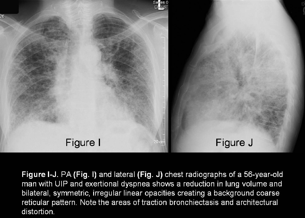
- Bilateral, symmetric, irregular linear opacities causing a reticular pattern (Fig. I and Fig. J)
- +/- concomitant ground-glass opacities, reduced lung volumes, and honeycombing
- Abnormalities may be diffuse; most commonly affect the lower lobes (80% cases) (Fig. I and J)
CT/HRCT
Early Findings
- Fine, irregular lines within the secondary pulmonary lobules (intralobular linear opacities)
- Irregular interlobular septal thickening
- Irregular pleural thickening
- Irregular interfaces between the lung parenchyma and the bronchovascular bundles
Typical Findings
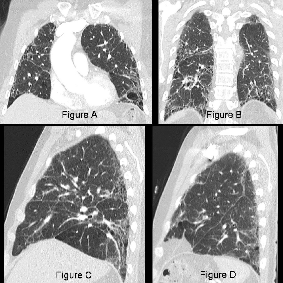
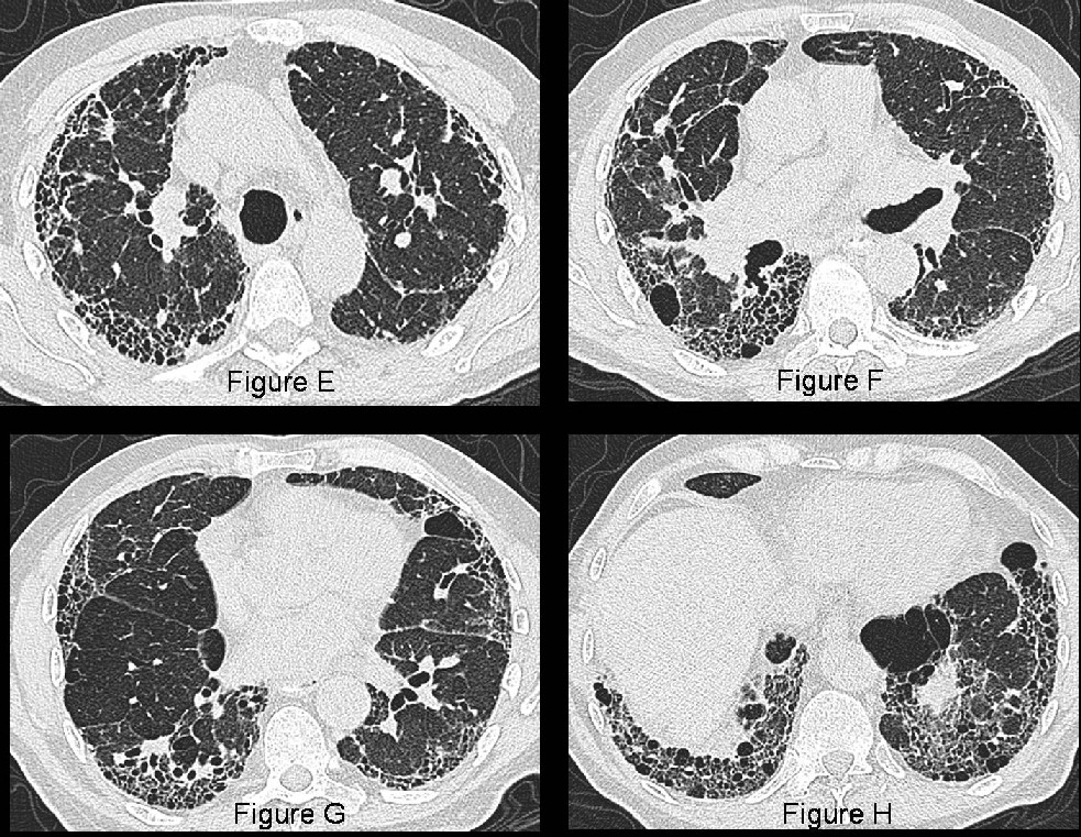
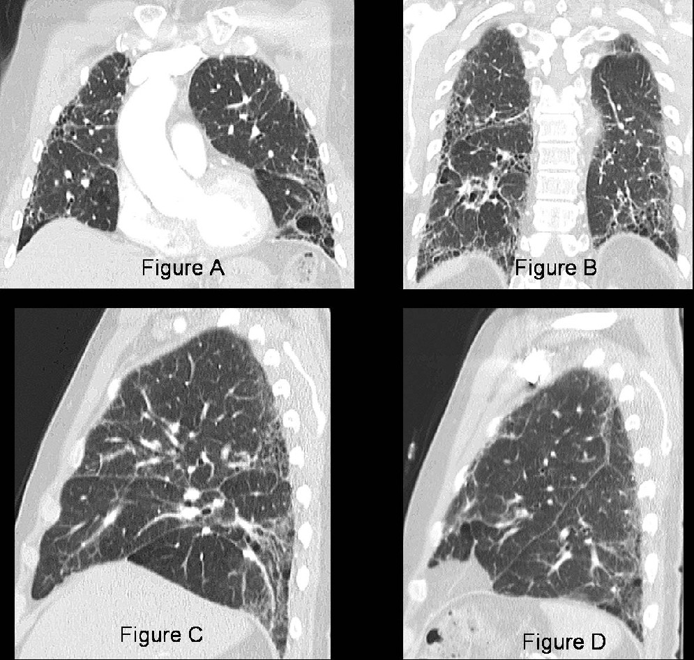
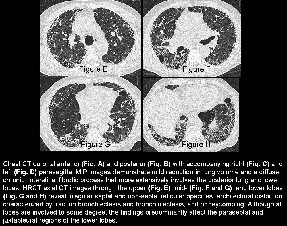
- Fibrosis and progressive lung parenchyma architectural distortion (Fig. A-H)
- Traction bronchiectasis and bronchiolectasis (Fig. A-H)
- Honeycombing (Fig. A-H)
- Predominance of paraseptal, peribronchiolar, basal and juxtapleural abnormalities (Fig. A-H)
- Reduction in lung volume
- Common mild mediastinal lymph node enlargement
- Most commonly involves the lower right paratracheal (Station 4R) and subcarinal (Station 7) lymph node stations
- Short axis diameters range between 10-21 mm
- More prevalent with more severe lung disease; associated with progression of fibrosis
- Foci of ground-glass attenuation correlate with the presence of active alveolitis (70% cases) or fibrosis beyond the resolution of the CT scan
Treatment
- Conventional management based on suppressing inflammation to prevent progression of ground-glass opacities to fibrosis with corticosteroids
- Aggressive immunosuppressive and cytotoxic treatment regimens have largely failed to reduce morbidity and mortality
- Lung transplantation in select patients
- Future therapies aimed at preventing or inhibiting the fibroproliferative response and promoting alveolar reepithelialization
Prognosis
Typical patient
- Slowly progressive course over years
- Mortality rate: 3.3 (men) and 2.5 (women) / 100,000 population
- Mean length of survival from time of diagnosis: 3.2-5.0 years
- Respiratory failure most common cause of death (40%)
- Other causes of death include: heart failure , bronchogenic carcinoma, pulmonary infection, cor pulmonale, ischemic heart disease, and pulmonary emboli
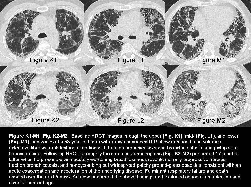
Accelerated UIP – IPF (Figure K1-M1; Fig. K2-M2)
- Small percentage of affected patients
- Accelerated decline or acute exacerbation of underlying disease
- Rapid downhill clinical course with acute marked increase in dyspnea and fulminant respiratory failure
- Usually refractory to therapy; very poor prognosis; death often within 1 week
- Characterized by new juxtapleural or patchy ground-glass and air space opacities on imaging (Fig. K2-M2)
Caveats
- Irregular linear opacities, honeycombing, and traction bronchiectasis indicate fibrosis and predict a poor response to therapy
- Combined with the clinical and radiographic findings, HRCT can render a high confidence diagnosis of UIP; obviating open lung biopsy in many cases
- Fibrosing NSIP can be difficult to differentiate from UIP. UIP tends to be primarily paraseptal, juxtapleural, or peribronchiolar in distribution. This pattern is less often seen with fibrosing NSIP. Accurate differentiation is based primarily on histology (i.e., fibrosing NSIP lacks temporal heterogeneity)
- An acute exacerbation or acceleration of the underlying disease process most often results in fulminant respiratory failure and death
Selected Readings
- Attili AK, Kazerooni EA, Gross BH, Flaherty KR, Martinez FJ. Thoracic lymph node enlargement in usual interstitial pneumonitis and nonspecific-interstitial pneumonitis: prevalence, correlation with disease activity and temporal evolution. J Thorac Imaging. 2006; 21(4):288-292.
- Bnà C, Zompatori M, Poletti V, Spaggiari E, Chetta A, Calabrò E, Ormitti F, Berti E, Cancellieri A, Chilosi M. Differential diagnosis between usual interstitial pneumonia (UIP) and nonspecific interstitial pneumonia (NSIP) assessed by high-resolution computed tomography (HRCT). Radiol Med. 2005; 109(5-6):472-487.
- Elliot TL, Lynch DA, Newell JD Jr, Cool C, Tuder R, Markopoulou K, Veve R, Brown KK. High-resolution computed tomography features of nonspecific interstitial pneumonia and usual interstitial pneumonia. J Comput Assist Tomogr. 2005; 29(3):339-345.
- Travis WD, Colby TV, Koss MN, Rosado de Christenson ML, Müller NL, King TE Jr. Diffuse parenchymal lung diseases. In: King DW, ed. Atlas of Nontumor Pathology: Non-Neoplastic Disorders of the Lower Respiratory Tract, first series, fascicle 2. Washington, DC: American Registry of Pathology; 2002: 59-73.
Original case written by its authors at Virginia Commonwealth University and published at this address as part of a weekly teaching collection. Reproduced here as an archive.