Diagnosis: Large partially obstructing esophageal epiphrenic diverticulum
Differential Diagnosis
- Benign obstructing esophageal lesions
- Stricture
- Achalasia
- Chagas Disease
- Malignant obstructing esophageal lesions
- Esophageal neoplasia
- Lymphadenopathy
- Esophageal dysmotility syndromes
- Scleroderma
- Hiatus Hernia
- Presbyesophagus
- Iatrogenic
- Esophagectomy with creation of a neoesophagus (e.g., gastric pull-through, colonic interposition)
- Esophageal speech prosthesis post laryngectomy
- Other
- Chronic inflammatory lung disease with adhesion formation
Background
Discussion
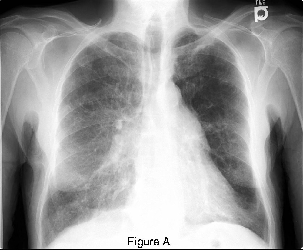
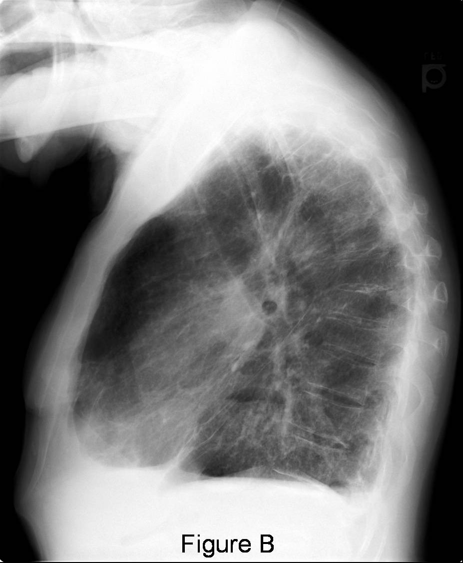
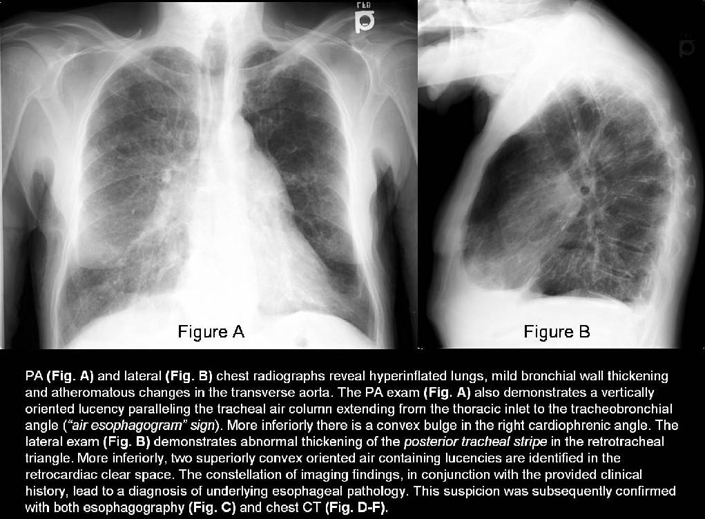
The “air esophogram” sign is created by an air-filled, non-collapsing esophagus signifying the presence of underlying intrinsic esophageal disease or chronic inflammatory pulmonary parenchymal disease with resultant adhesions between the lung and esophagus. The posterior tracheal stripe is a vertical stripe seen on lateral chest radiography formed by air within the trachea and right lung outlining the posterior tracheal wall and intervening soft tissues. It typically measures 2.5 mm in thickness. However, when the posterior trachea comes in contact with the anterior wall of the esophagus, the esophageal wall, the posterior tracheal wall, and intervening soft tissues combine to form a thicker tracheoesophageal stripe that measures up to 5.5 mm. More significant thickening of the posterior tracheal stripe occurs in the setting of various esophageal lesions, lymphatic malformations, mediastinitis, post-traumatic hematomas, mantle radiation therapy, and acquired vascular lesions. Right cardiophrenic angle opacities may result from dilatation of the right atrium, aneurysms of the ventricle or sinus of Valsalva; epicardial fat pad; pericardial cyst; Morgagni hernia; lymphadenopathy; paraesophageal varices; primary lung lesions; hiatus hernia; and epiphrenic diverticula. Epiphrenic diverticula are rare and only comprise 10% of all esophageal diverticula. The majority occur in the middle aged and elderly. Epiphrenic diverticula are further categorized as pulsion diverticula and are considered pseudodiverticula. That is, only the mucosa and a variable amount of submucosa herniate through the muscularis propria as a result of increased intraluminal pressure. The majority occur in the distal 10 cm of the esophagus, usually along the right lateral wall.
Etiology
The etiology of epiphrenic diverticula is not fully understood. Esophageal obstruction, motility disorders and weakness of the esophageal wall are believed to be underlying predisposing factors. Congenital epiphrenic diverticula exist but are exceedingly rare.
Clinical Findings
Many patients are asymptomatic and are incidentally diagnosed when radiologic studies are performed for other reasons. Symptoms are more likely to occur as the size of the diverticulum increases. Dysphagia, regurgitation and nocturnal cough are the more common presenting symptoms.
Imaging Findings
Radiography
- Frontal exam may reveal a convex bulge along in either the right or left cardiophrenic angle (Fig. A)
- “Air esophogram” sign may be present (Fig. A)
- Lateral exam may reveal a thickened posterior tracheal stripe (Fig. B) and or a large soft tissue mass mimicking a hiatus hernia.
- Retrocardiac air-fluid level(s) may be present (Fig. B)
CT
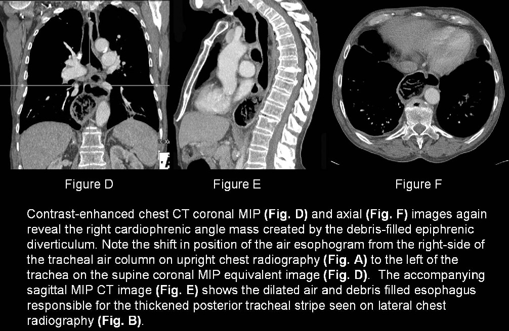
- May demonstrate retained esophageal debris (Fig. D-F) or air-fluid level in esophagus
- “Air esophogram” sign may be present (Fig. D and E)
- Thin walled, air or air and fluid filled structure communicating with the esophagus (Fig. D-F)
Barium esophogram
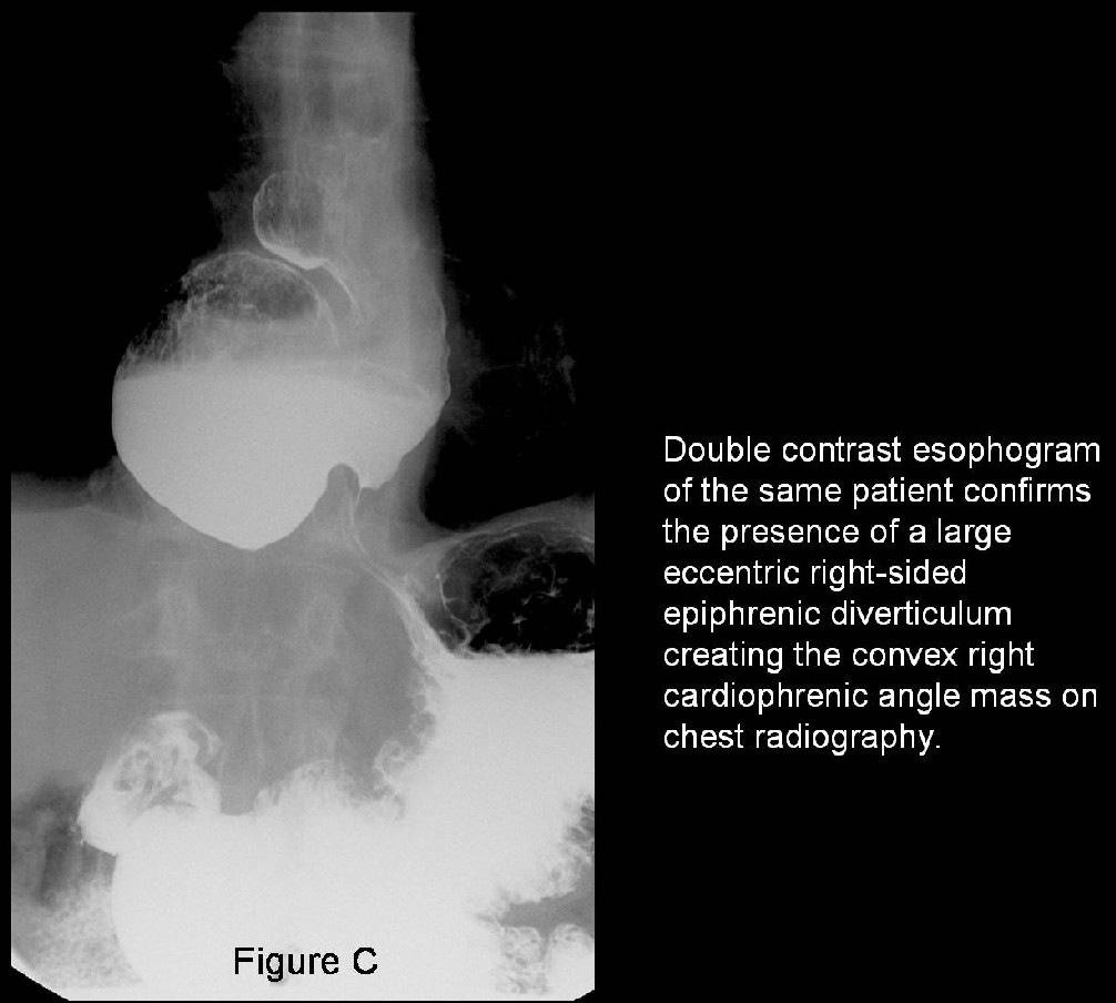
- May reveal a barium filled sac with rounded contour in the distal esophagus (Fig. C)
- Features of achalasia and hiatus hernia may be seen and are often associated with epiphrenic diverticula
Management
- Asymptomatic patients do not require surgery.
- Symptomatic patients
- Diverticulectomy and myotomy
Selected Readings
- Brant WE, Helms, CA. Pharynx and Esophagus. Fundamentals of Diagnostic Radiology. Vol II., 3rd ed. Philadelphia: Lippincott, Williams, & Wilkins. 2007; 805.
- Duda M et al. Etiopathogenesis and classification of esophageal diverticula. Int Surg. 70(4):291-5, 1985.
- Gibbs, JM, Chandrasekhar CA, Ferguson EC, Oldham SAA. Lines and Stripes: Where did they go? –From conventional radiography to CT. RadioGraphics 2007; 27: 33-48.
- Jordan PH Jr, Kinner BM. New look at epiphrenic diverticula. World J Surg 1999; 23: 147-52.
- Proto AV, Lane EJ. Air in the esophagus. A frequent radiographic finding. AJR 1977; 129:433-440.
Original case written by its authors at Virginia Commonwealth University and published at this address as part of a weekly teaching collection. Reproduced here as an archive.