Diagnosis: Nummular sarcoidosis
Differential Diagnosis:
- Bronchoalveolar carcinoma or other primary lung cancers
- Pulmonary metastases
- Granulomatous diseases (Mycobacterium tuberculosis, non-tuberculous mycobacterial infections, bronchopulmonary fungal infections)
- Wegener granulomatosis
- Septic emboli
- Kaposi sarcoma
Discussion
Background
Sarcoidosis is a systemic granulomatous disease characterized by the presence of non-caseating granulomas and proliferation of epithelioid cells. It affects primarily the lungs and lymphatic system. Nummular, or nodular, sarcoidosis is an uncommon imaging manifestation of pulmonary sarcoidosis. The prevalence of this particular form of sarcoidosis is estimated to be approximately 2.4 – 4%.
Etiology
Although no clear cause has been established, one postulated theory includes an abnormal immune response to an unidentified antigen. Increased risk in persons with family histories of the disorder suggests a yet unidentified genetic predisposition.
Clinical Findings
The peak incidence of sarcoidosis occurs in African American females in the third decade of life; however, it may affect persons of any age, gender, or race. The annual incidence of sarcoidosis in the United States is estimated at 35.5 per 100,000 for African Americans and 10.9 per 100,000 for Caucasians. Nodular or nummular sarcoidosis often demonstrates a similar epidemiological distribution. The degree of extrapulmonary involvement is similar to other forms of sarcoidosis, and can include the lymph nodes, skin, eyes, kidneys, heart, liver, salivary glands, sinuses, muscles, bones, and central nervous system. Diagnosis is definitively made by bronchoscopy with transbronchial biopsy.
Pathology
- Well-formed, non-caseating epithelioid granulomas in a perilymphatic or bronchocentric distribution.
Imaging Findings
Radiography
Parenchymal involvement
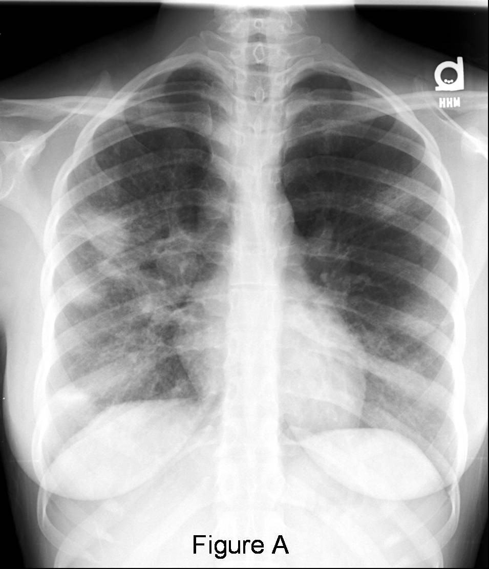
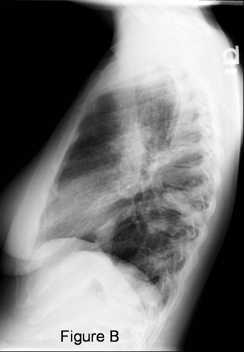
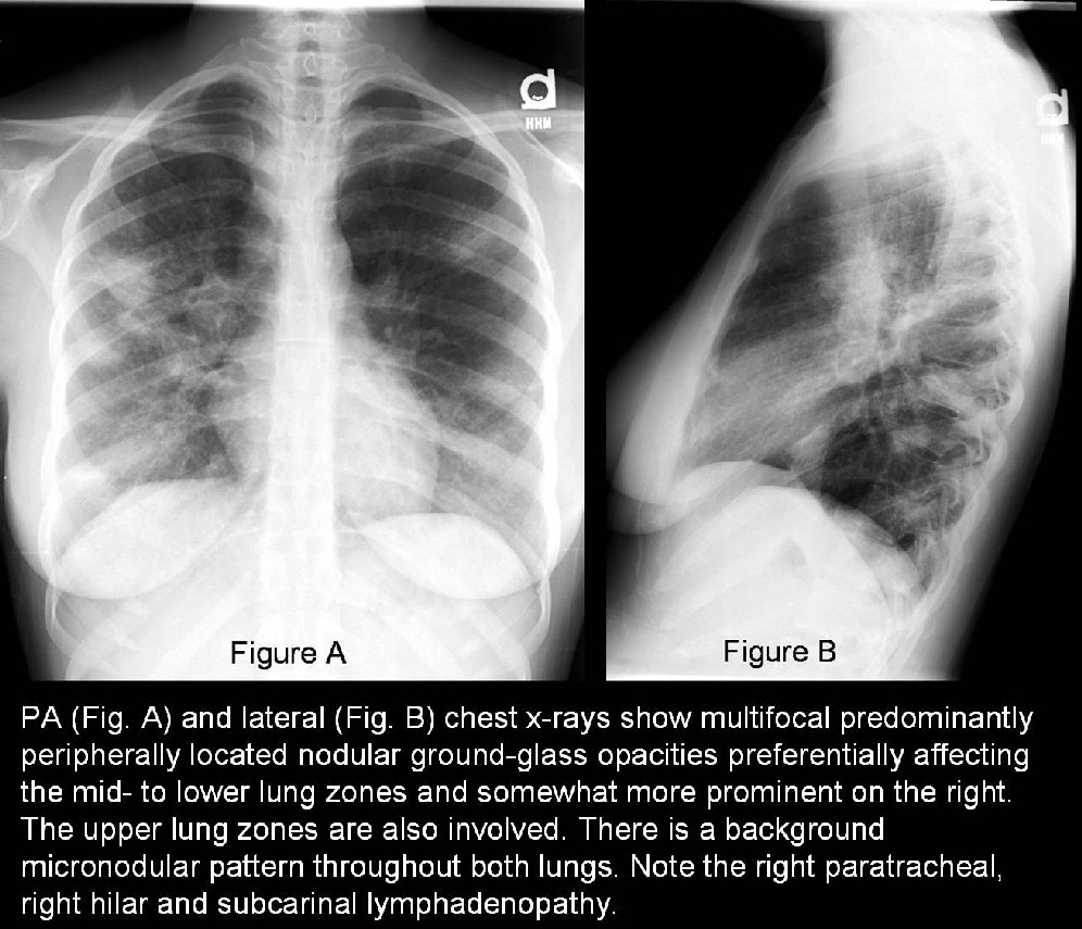
- Nummular (aka nodular or alveolar) sarcoidosis presents with single or multiple nodules (<3cm) or masses (>3cm) with or without air bronchograms (Fig. A and Fig. B)
- Small bilateral reticular or nodular opacities; upper or middle lung zones (Fig. A and Fig. B)
- Rarely focal opacity, pleural effusion, pneumothorax, cavitation
- Fibrosis with upper lobe volume loss; hilar retraction; cystic or honeycomb changes; complicating mycetoma
Intrathoracic Lymphadenopathy
- A classic triad of bilateral hilar and right paratracheal lymphadenopathy is common.
- May see varying degrees of hilar, mediastinal, or paratracheal lymphadenopathy (Fig. A and Fig. B); possibly with peripheral calcifications of lymph nodes
CT/HRCT
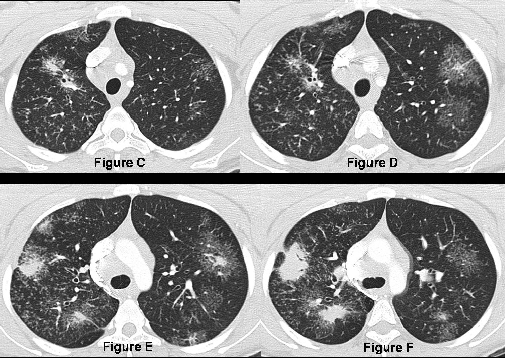
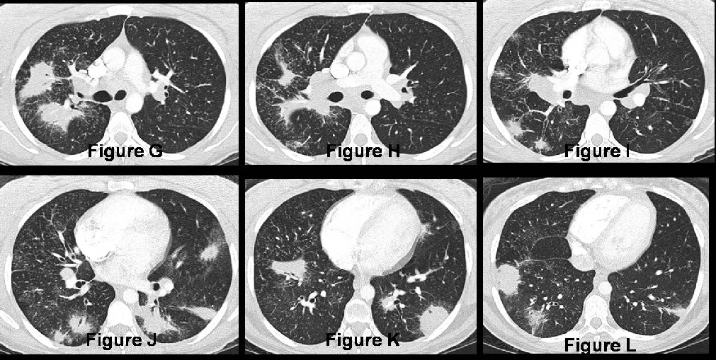
- Nodular sarcoidosis – single or multiple pulmonary nodules/masses in a perilymphatic distribution, often with air bronchograms; nodules may be surrounded by ground glass opacities (Fig. C-L)
- Classic sarcoidosis –
- Discrete or irregular, 1 to 5 mm nodules in a perilymphatic or bronchovascular distribution (Fig. C-L)
- Irregular septal thickening with a beaded appearance (Fig. C-L)
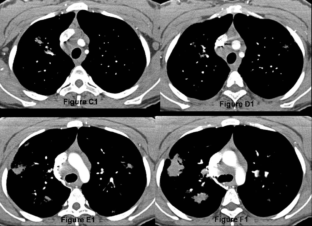
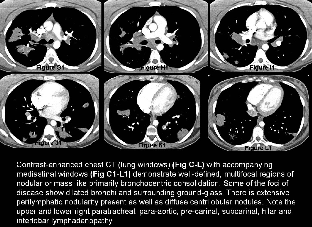
- Lymphadenopathy (Fig. C1-L1)
- Superior hilar displacement
- Pulmonary fibrosis: central and upper lobe distribution; architectural distortion; traction bronchiectasis; and large cystic spaces
Treatment and Prognosis
- Spontaneous remission occurs in nearly two thirds of patients.
- Corticosteroids are the first line of treatment, although the optimal dose and duration is not well documented.
- The prognosis of patients with nodular sarcoidosis is favorable, and complete resolution may be seen.
Suggested Readings
- Brant WE, Helms, CA. Diffuse Lung Disease. Fundamentals of Diagnostic Radiology. Vol II., 3rd ed. Philadelphia: Lippincott, Williams, & Wilkins. 2007; 500-504.
- Collins J., Stern EJ. Upper Lung Disease, Infection, and Immunity. Chest Radiology: The Essentials.-2nd ed. Philadelphia: Lippincott, Williams, & Wilkins. 2008; 165-168.
- Malaisamy S, et al. The Clinical and Radiologic Features of Nodular Pulmonary Sarcoidosis. Lung. 2009; 187:9-15.
- Park HJ, et al. Typical and Atypical Manifestations of Intrathoracic Sarcoidosis. Korean J Radiol; 10:623-631.
- Parker MS, Rosado de Christenson, Abbott GF. Diffuse Lung Disease: Case 127 Sarcoidosis. In: Teaching Atlas of Chest Imaging. New York: Thieme Medical Publishers, Inc. 2006; 485-489.
Original case written by its authors at Virginia Commonwealth University and published at this address as part of a weekly teaching collection. Reproduced here as an archive.