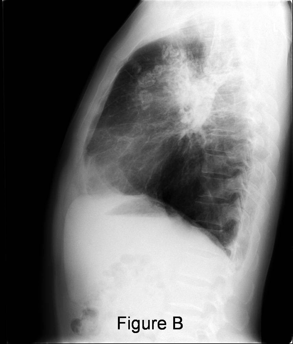
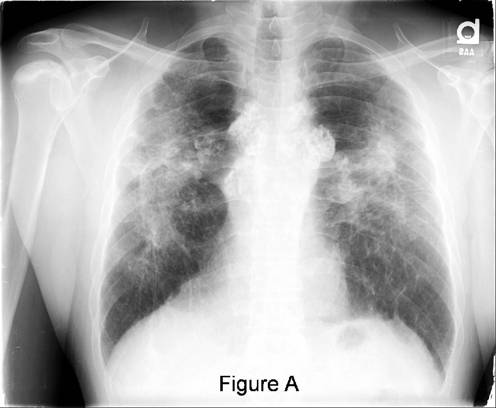
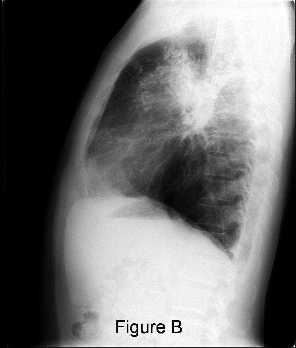
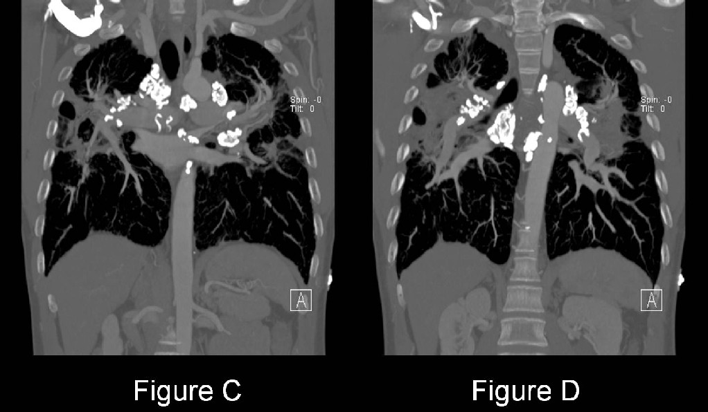
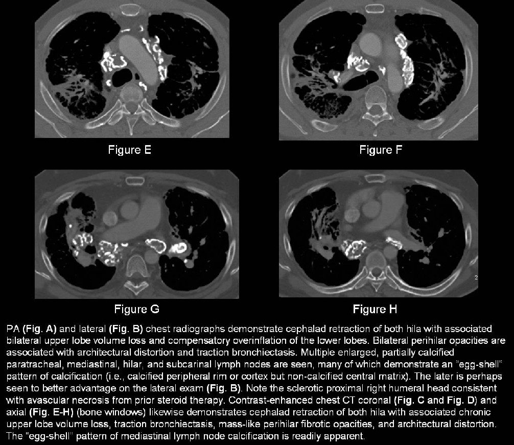
Diagnosis: Stage 4 Sarcoidosis with “egg-shell” pattern of lymph node calcifications
Differential Diagnosis:
- Complicated silicosis
- Coal worker’s pneumoconiosis
- Granulomatous diseases (Mycobacterium tuberculosis, various pulmonary fungal infections)
- Treated lymphoma
Discussion
Background
Sarcoidosis is a common systemic granulomatous disease that typically affects the lung and the lymphatic system.
Etiology
Although no clear cause has been established, an abnormal response of the immune system to an unidentified antigen is postulated to play a role it is pathogenesis. There may also be an underlying genetic predisposition in some reported familial cases.
Clinical Findings
Sarcoidosis is seen in persons of all ages and both sexes, however, it classically presents in African American women between the ages of 20 and 40. The annual incidence of sarcoidosis in the United States is estimated at 35.5 per 100,000 for African Americans and 10.9 per 100,000 for Caucasians. Sarcoidosis most commonly involves the lungs and lymph nodes; however, it can also involve the skin, eyes, kidneys, heart, liver, salivary glands, sinuses, muscles, bones, and central nervous system. Most often, affected patients present with symptoms of dry cough, dyspnea, and chest pain. However, constitutional symptoms, including palpable lymphadenopathy and skin lesions are also quite common. Sarcoidosis is also associated with certain syndromes such as Heerfordt syndrome (e.g., fever, parotid enlargement, facial palsy, anterior uveitis) and Löfgren syndrome (e.g., acute sarcoidosis with fever, polyarthralgias, erythema nodosum, and bilateral hilar lymphadenopathy). Approximately 20% of patients with sarcoidosis develop pulmonary fibrosis with pulmonary insufficiency and pulmonary artery hypertension. Some asymptomatic patients are diagnosed incidentally on radiographs obtained for other purposes.
Pathology
- Well-formed, non-caseating epithelioid granulomas are the hallmark
Imaging Findings
Conventional Chest Radiographic Staging
Stage 0: No radiographic abnormality
Stage 1: Hilar and or mediastinal lymphadenopathy
Stage 2: Lymphadenopathy with evidence of parenchymal involvement
Stage 3: Parenchymal involvement; no visible lymphadenopathy
Stage 4: Upper lobe fibrosis (Fig. A and Fig. B)
- Hilar retraction
- Volume loss
- Elevated diaphragm
- Cystic changes
Radiography
Intrathoracic Lymphadenopathy
Garland triad
- Bilateral hilar lymphadenopathy and right paratracheal lymphadenopathy (also called the 1,2,3 sign) (Fig. A and Fig. B)
- May see varying degrees of hilar, mediastinal, or paratracheal lymphadenopathy +/- “eggshell pattern” of calcifications (Fig. A and Fig. B)
Parenchymal involvement
- Usually consists of small bilateral reticular or nodular opacities; upper or middle lung zones.
- Nodular (i.e., alveolar sarcoidosis): multifocal nodules or masses with or without air bronchograms
- Rarely focal opacity, pleural effusion, pneumothorax, cavitation
- Fibrosis with upper lobe volume loss; hilar retraction (Fig. A and Fig. B); cystic or honeycomb changes; complicating mycetoma
CT/HRCT
- Discrete or irregular, 1- to 5- mm nodules in a perilymphatic and or bronchovascular distribution; involvement of the juxtapleural surface, interlobular septa, and secondary pulmonary lobules.
- Ground-glass opacities, large nodules, and masses in a perilymphatic distribution
- Irregular septal thickening with a beaded appearance.
- Pulmonary fibrosis: central and upper lobe distribution; architectural distortion; traction bronchiectasis; and large cystic spaces (Fig. C-H)
- Conglomerate central masses with superior hilar displacement (Fig. C-H)
- Peripheral honeycombing, bullae, cystic spaces complicated by mycetoma
Scintigraphy
Gallium-67 uptake patterns suggestive of sarcoidosis
- Lambda pattern: homogeneous perihilar, infrahilar, paratracheal lymph node uptake
- Panda pattern: bilateral symmetrical lacrimal gland and parotid gland uptake
Treatment
- 60-80% of patients: self-limiting course of disease and requires no specific intervention.
- Most treatment is based on treating symptoms, usually with corticosteroids to reduce inflammation.
- While this treatment appears to work in the short term, long term benefits are not clear.
- Additional treatment options for patients who are steroid resistant or intolerant include
- Methotrexate
- Azathioprine
- Cyclophosphamide
- Chloroquinine
Prognosis
- 60-80% of patients: Self-limiting course with no long term morbidity
- 20-30% of patients: Some permanent lung damage will occur
- 10-15% of patients: Develop a chronic form of the disease
- 5-10 of patients: Life-threatening complications with severe pulmonary or other organ system compromise
Suggested Readings
- Brant WE, Helms, CA. Diffuse Lung Disease. In: Fundamentals of Diagnostic Radiology. Vol II., 3rd ed. Philadelphia: Lippincott, Williams, & Wilkins. 2007; 500-504.
- Chiles C. Imaging features of thoracic sarcoidosis. Semin Roentgenol 2002; 37:82-93.
- Collins J., Stern EJ. Upper Lung Disease, Infection, and Immunity. In: Chest Radiology: The Essentials.-2nd ed. Philadelphia: Lippincott, Williams, & Wilkins. 2008; 165-168.
- Koyama T et al. Radiologic Manifestations of Sarcoidosis in Various Organs. RadioGraphics.2004; 24:87.
- Parker MS, Abbott GF. Diffuse Parenchymal Lung Disease. In: Teaching Atlas of Chest Imaging. New York: Thieme Medical Publishers, Inc. 2006; 485-489.
Original case written by its authors at Virginia Commonwealth University and published at this address as part of a weekly teaching collection. Reproduced here as an archive.