Diagnosis: Right atrial myxoma
Differential Diagnosis
- Other Primary Cardiac Tumors
- Secondary Cardiac Tumors
- Atrial Thrombus
Discussion
Background
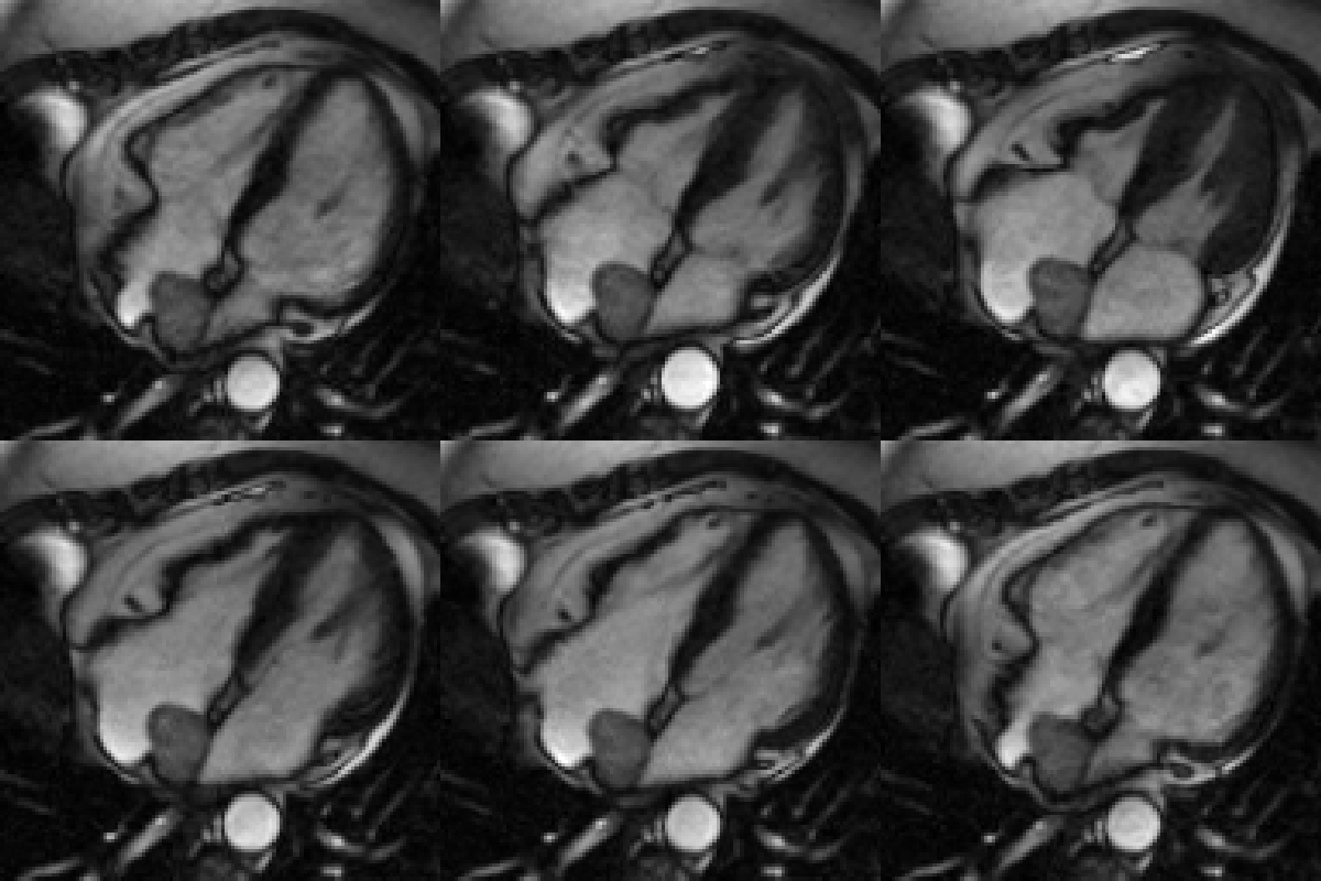
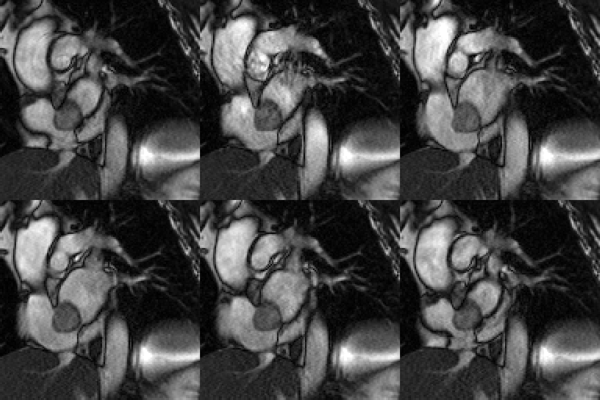
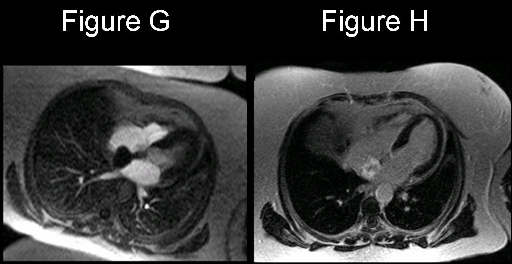
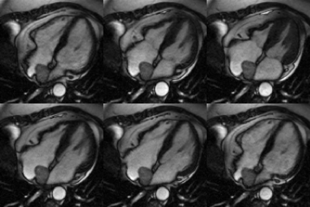
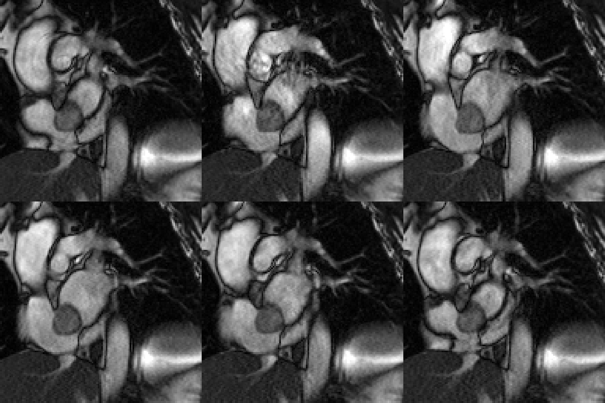
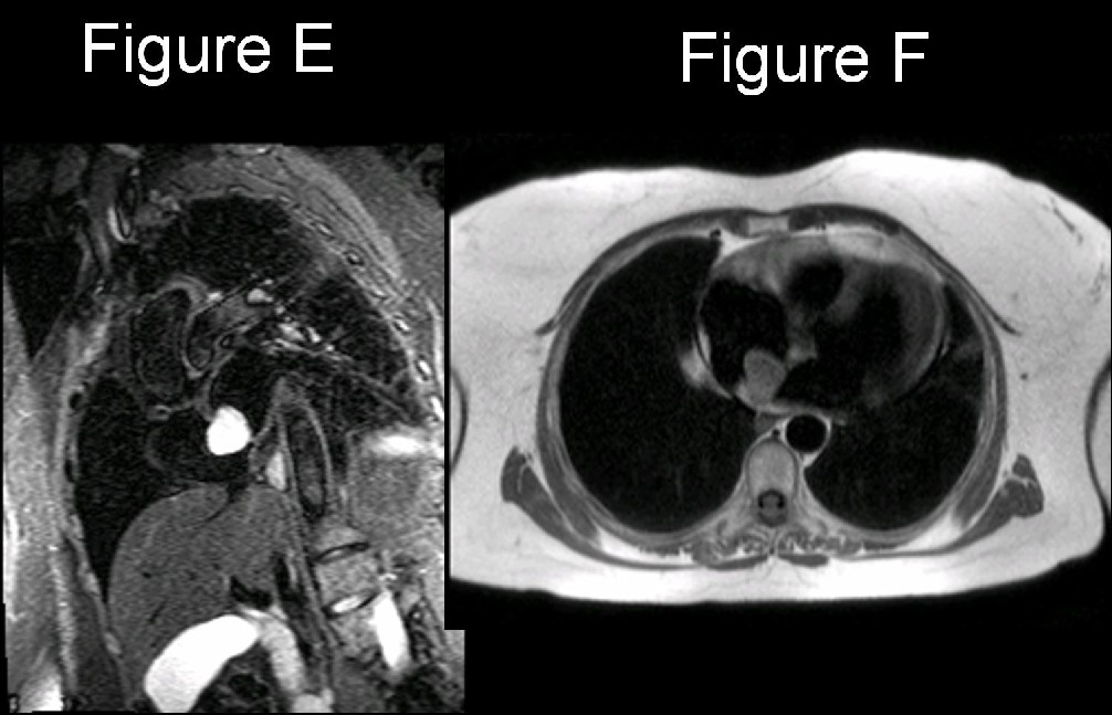
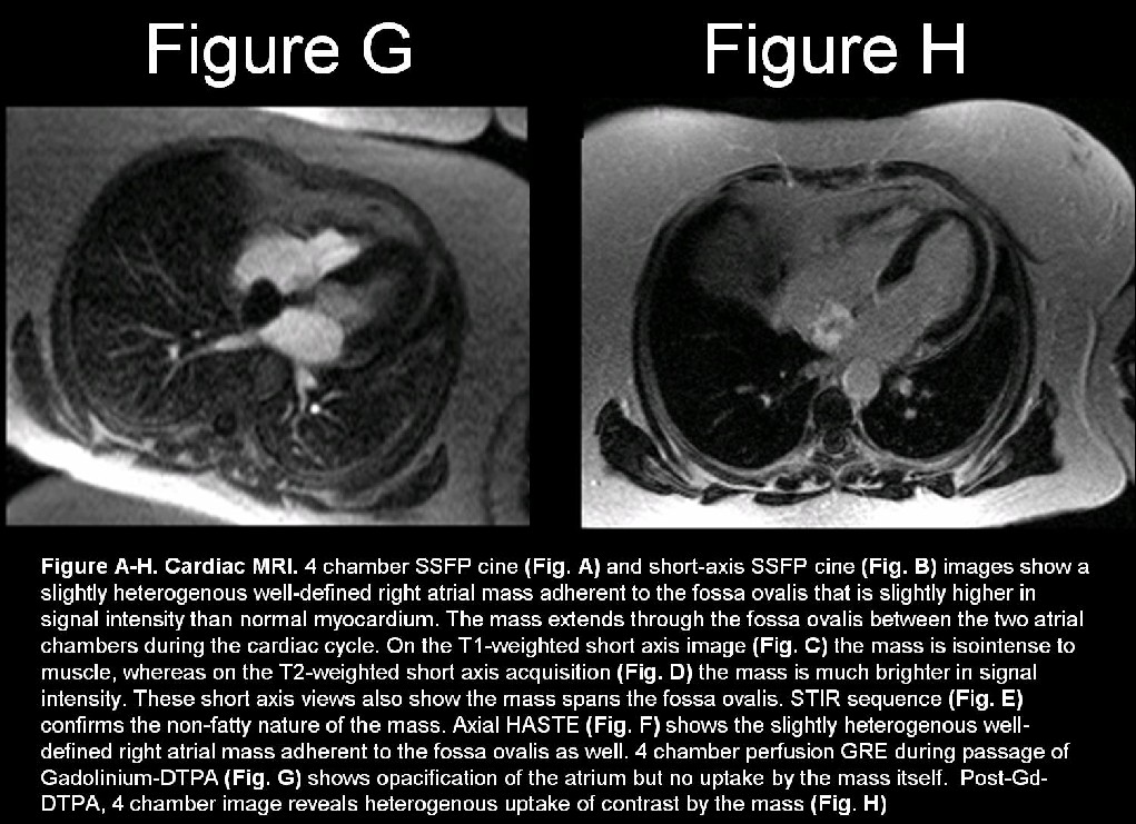
Secondary cardiac masses are 20-40 times more common than primary cardiac masses. Whereas most secondary cardiac masses are malignant, most primary cardiac masses are benign. Myxoma is the most common primary benign cardiac mass. Approximately 90% of myxomas are solitary pedunculated masses but as many as 5% may present as multiple masses. These relatively gelatinous tumors most often arise in close proximity to the fossa ovalis. Nearly 75- 85% originate in the left atrium; up to 25% occur in the right atrium and/or extend through the fossa ovalis (Fig. A-H); and about 5% arise within the ventricles. Multiple myxomas may be associated with various syndromes, including Carney syndrome (e.g., atrial myxomas, melanotic schwannomas, Cushing syndrome, multiple cerebral fusiform aneurysms, and breast fibroadenomas).
Etiology
Most cases of atrial myxoma are sporadic. Approximately 10% of myxomas may be inherited in an autosomal dominant manner. Multiple tumors occur in approximately 50% of familial cases and in these later cases are more frequently located in the ventricle.
Clinical Findings
Approximately 75% of sporadic myxomas occur in females. However, the female sexual predilection is less pronounced in familial atrial myxomas. The mean age at presentation for sporadic cases is 56 years, whereas the mean age for familial myxoma is 25 years. Most patients with sporadic myxomas are symptomatic and most commonly present with dyspnea. In about 20% of cases, the myxoma does not cause symptoms and is incidentally discovered. Symptoms are produced by mechanical cardiac chamber or valvular obstruction or tumor embolization. Tumor embolism occurs in 30-40% of patients. The site of embolism depends upon whether the myxoma is located in the left or right atrium. The symptoms and signs of left atrial myxomas often mimic mitral stenosis. Right atrial myxomas grow to approximately twice the size of typical left atrial myxomas before becoming symptomatic, and are sometimes associated with tricuspid stenosis and atrial fibrillation. Symptoms may be precipitated by a change of body position and may include: orthopnea; difficulty breathing when asleep; chest pain; syncope or near-syncope; and palpitations. Constitutional symptoms are observed in 50% of patients and include: fever, weight loss, arthralgias, and Raynaud phenomenon, possibly related to the overproduction of interleukin-6. Hemoptysis due to pulmonary edema or infarction is seen in up to 15% of patients. Abnormal heart sounds including a “tumor plop” may be heard during auscultation when the tumor changes position as the patient changes position.
Imaging Findings
Radiography
- Enlarged left atrial appendage and enlarged left atrium; mimicking mitral stenosis
- Enlarged pulmonary veins and cephalization of blood flow; mimicking mitral stenosis
- +/- Unusual intracardiac tumor calcification
Transthoracic Echocardiography (TTE)
- Assess tumor location, size, attachment, and mobility
- Doppler can show the hemodynamic consequences of atrial myxoma consistent with mitral stenosis or regurgitation
Transesophageal echocardiography (TEE)
- Better resolution, specificity and 100% sensitivity compared to TTE
- Better visualization of tumor morphology, size, and the presence of a stalk
CT
May be useful in differentiating atrial myxoma from intracardiac thrombus:
Atrial Myxoma
- Attenuation: 43 ± 14 HU
- Larger than thrombus; 33 ± 16 mm
- Unenhanced scans: lower in attenuation than atrial blood
- Contrast-enhanced scans: heterogeneous mass (reflecting hemorrhage, necrosis, cyst formation, fibrosis, calcification)
- Location: in or near fossa ovalis
- Shape: smooth to lobular, often with polypoid projections
- Mobility: may demonstrate prolapse across the fossa ovalis or into the ventricular cavity depending on length of the peduncle.
- May demonstrate calcification(s)
Intracardiac Thrombus
- Attenuation: 57 ± 30 HU
- Smaller than myxomas; 21 ± 7 mm
- Homogenous more commonly but may appear heterogeneous
- Location: Posterior or lateral wall of atrium; atrial appendage
- Shape: Flat, smooth, lobulated or pedunculated
- May demonstrate calcification(s)
MR
Site of attachment better visualized with a post-surgical correlation of 83%.
Cine MR gradient echo (GRE) images can demonstrate the mobility of a tumor.
Useful in differentiating atrial myxoma from intracardiac thrombus:
Atrial Myxoma
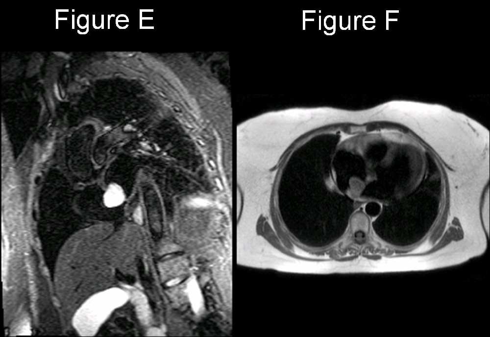
- HASTE: heterogenous mass; signal intensity slightly higher than normal myocardium (Fig. F)
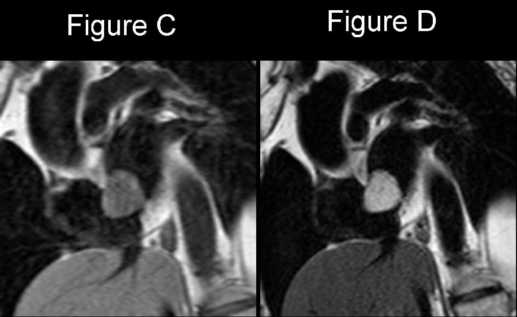
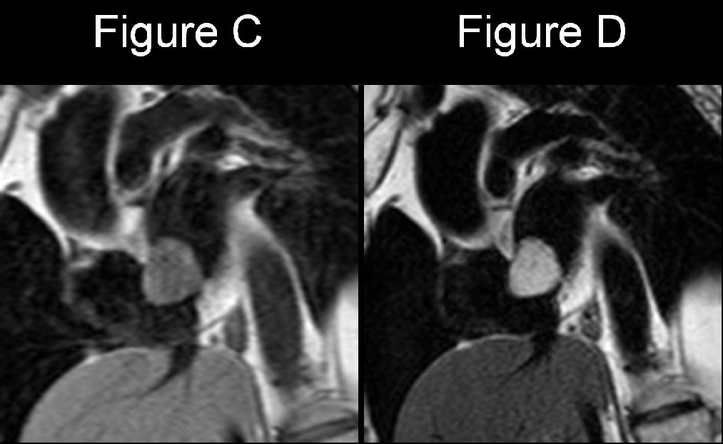
- T1WI: mass isointense relative to myocardium (Fig. C)
- T2WI: mass ↑ SI relative to myocardium (Fig. D)
- STIR: mass demonstrates no fat suppression (Fig. E)
- SSFP cine sequence: ↑ SI (T2/T1 ratio) secondary to gelatinous composition of tumor with a high fluid-like content (relative T2-weighting) (Fig. A; B)
- Cine images: pedunculated mass may prolapse across fossa ovalis or atrial-ventricular valve
- Perfusion GRE: no uptake of Gd-DTPA by the mass (Fig. G)
- Post-Gd-DTPA: heterogenous enhancement of mass possible (Fig. H)
Intracardiac Thrombus
- Location: usually atrial appendage; broad-based; irregular contour; may appear layered
- Thrombus adherent to cardiac chamber walls are akinetic on cine acquisitions
- SSFP sequence: ↔ / ↓ SI relative to normal myocardium
- Post-Gd-DTPA: no to mild heterogenous enhancement
- Signal intensity characteristics vary depending on acuteness or chronicity on the thrombus:
T1 PD T2 GRE HASTE Acute Thrombus ↔ ↔ ↔ / ↑ ↔ ↔ Chronic Thrombus ↔ ↔ / ↓ ↔ / ↓ ↓ / ↓↓
Treatment
- Surgical resection of the myxoma is the treatment of choice.
- Damaged valves may require annuloplasty or prosthetic replacement.
Prognosis
- Surgery for sporadic atrial myxoma is usually curative.
- Long-term prognosis is excellent.
- Recurrence rate is 1-5%.
- Recurrence after 4 years is uncommon.
- Recurrence rate for familial atrial myxoma is 20%.
- Untreated myxoma can be complicated by systemic embolization.
- Sudden death may occur in up to 15% patients with untreated atrial myxoma.
- Secondary to coronary or systemic embolization
- Obstruction of blood flow at the mitral or tricuspid valve.
Caveats
- MRI is more sensitive than echosonography or CT for differentiating atrial myxoma from atrial thrombus. Thrombus is usually situated in the posterior wall of the atrium and has a layered appearance. The presence of a stalk and mobility favors atrial myxoma.
- On CT, atrial myxomas and thrombi can sometimes be differentiated by their distinguishing features of size, origin, shape, mobility, and prolapse. Attenuation coefficients and or the presence of calcification are not useful discriminating features.
Suggested Readings
- Araoz, P.A., et al., CT and MR imaging of benign primary cardiac neoplasms with echocardiographic correlation. RadioGraphics 2000; 20(5): 1303-1319.
- Grebenc ML, Rosado-de-Christenson ML, Green CE, Burke AP, Galvin JR. Cardiac myxoma: imaging features in 83 patients. RadioGraphics 2002; 22(3):673-689.
- Grizzard JD, Judd RM, Kim RJ. Cardiovascular MRI in Practice: A Teaching File Approach. Springer-Verlag, London Limited 2008; 262-263.
- Hoffmann, U., et al., Usefulness of magnetic resonance imaging of cardiac and paracardiac masses. Am J Cardiol. 2003; 92(7): 890-895.
- Scheffel H, Baumueller S, Stolzmann P, Leschka S, Plass A, Alkadhi H, Schertler T. Atrial Myxomas and Thrombi: Comparison of Imaging Features on CT. AJR 2009; 192:639-645.
- Wintersperger, B.J., et al. Tumors of the cardiac valves: imaging findings in magnetic resonance imaging, electron beam computed tomography, and echocardiography. Eur Radiol 2000; 10(3): 443-449.
Original case written by its authors at Virginia Commonwealth University and published at this address as part of a weekly teaching collection. Reproduced here as an archive.