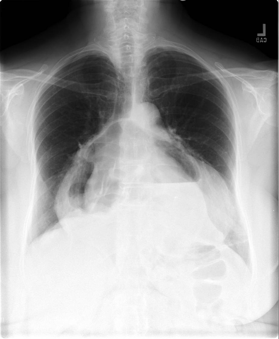
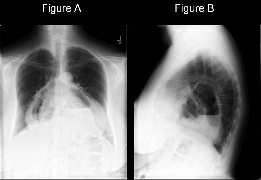
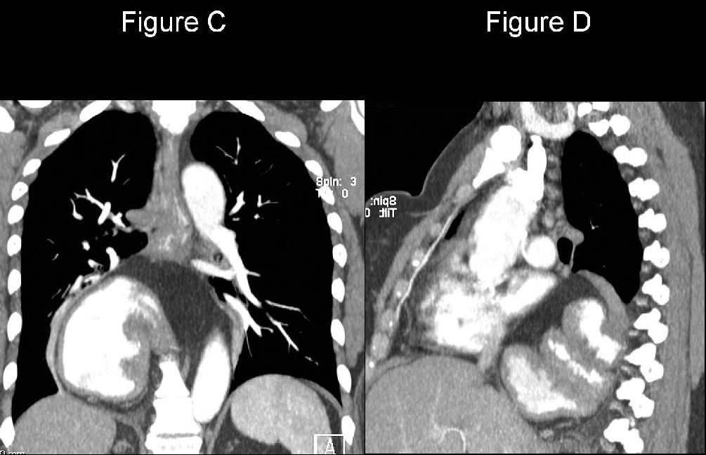
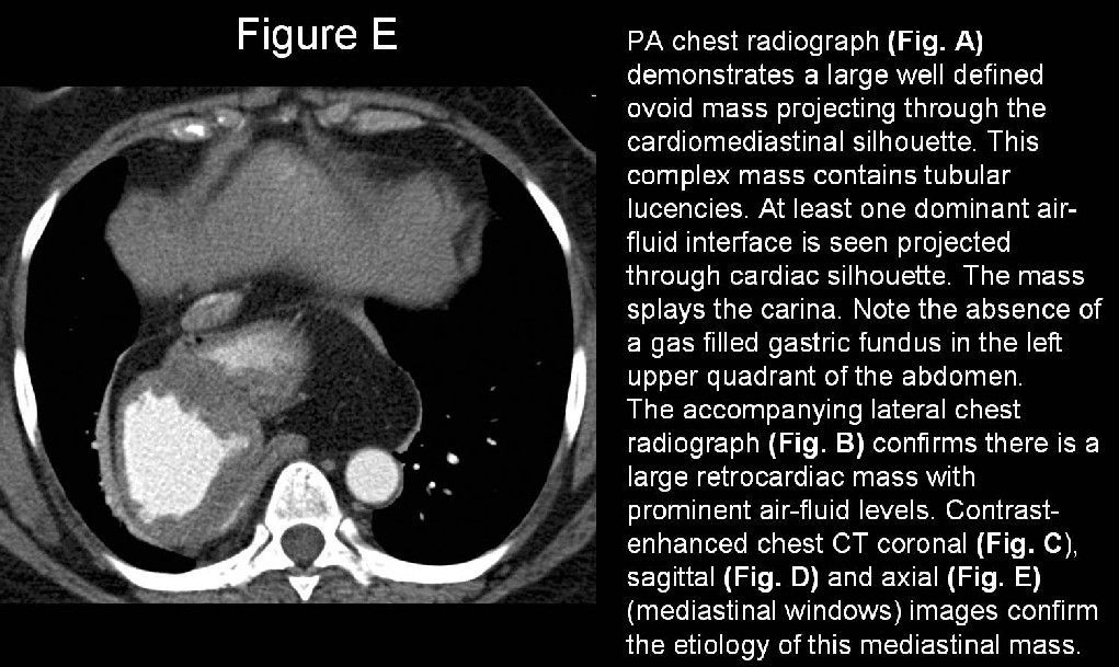
Diagnosis: Hiatus Hernia
Differential Diagnosis
- Pneumopericardium
- Bochdalek Hernia
- Morgagni Hernia
Discussion
Background
Hiatus hernia represents an anatomical abnormality in which part of the stomach prolapses through the diaphragm and into the mediastinum. Hiatus hernia is present in approximately 15% of the population, but is associated with symptoms in only a minority of those persons.
Etiology
Hiatus Hernia: Most likely results from a capacious diaphragmatic esophageal hiatus allowing the stomach to “slip and slide” through the opening (hence the lay term sliding hernia) into the thorax. Additional potential contributors to hiatus hernia may include a shortened esophagus caused by inflammation and scarring from reflux disease which pulls the stomach cephalad and an abnormally loose attachment of the esophagus to the diaphragm subsequently allowing prolapse of the esophagus and stomach.
Bochdalek Hernia: Congenital defect in the foramen of Bochdalek (pleuroperitoneal hiatus) of the diaphragm that allows various intra-abdominal organs to prolapse into the thoracic cavity. Most such defects occur in the posterior lateral left diaphragm.
Morgagni Hernia: Rare defect of the anterior medial diaphragm associated with a retrosternal, or parasternal hernia and responsible for approximately 2% of all congenital diaphragmatic hernias. The majority of these hernias occur on the right side of the body. Most affected patients are asymptomatic.
Pneumopericardium: Pneumopericardium may result from cardiac or mediastinal surgery, penetrating or blunt trauma, infectious pericarditis with gas-producing organisms, and a fistulous communication between the pericardium and an adjacent air-containing organ (i.e. stomach or esophagus). Pneumopericardium secondary to blunt chest trauma is generally due to 1 of 3 mechanisms: (1) air extension along pulmonary venous perivascular sheaths from ruptured alveoli to the pericardium; (2) pneumothorax associated with a pleuropericardial tear; or (3) a direct tracheobronchial–pericardial communication.
Clinical Findings
Non-traumatic hiatus hernia is usually related to obesity or pregnancy. The larger the hernia, the more likely it is to cause symptoms. The most common symptom is gastroesophageal reflux. Although hiatus hernia may contribute to reflux disease, it is unclear if it alone results in reflux disease. Larger hernias may compress adjacent mediastinal structures and airways resulting in various signs and symptoms as well such as dysphagia, wheezing, and atelectasis.
Imaging Findings
Esophagography
Hiatus hernia:
- Sliding hiatus hernia:
- Entire gastroesophageal junction (GEJ) and a variable portion of the stomach prolapse above the diaphragm.
- Paraesophageal hernia:
- GEJ remains below the diaphragm and a portion of the gastric fundus herniates into the thoracic cavity adjacent to the esophagus.
- Massive hiatus hernia:
- Large diaphragmatic defect allows herniation of a large amount of abdominal viscera into the thoracic cavity.
Radiography / CT
- Bochdalek hernia:
- Focal posteromedial mass which may be unilateral or bilateral.
- May contain retroperitoneal fat, kidney, or air containing bowel loops.
- Morgagni hernia:
- Small focal anteromedial mass
- Generally lateral or just posterior to the xyphoid process.
- Most commonly unilateral and right-sided.
- Pneumopericardium:
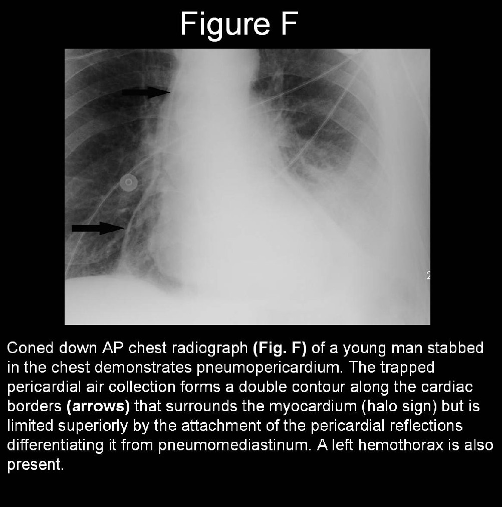
- “Halo” sign- radiolucent band of air partially or completely surrounds the heart, does not extend above the upper limit of the pericardial reflections, and is confined to below the aortic arch (Fig. F)
- “Transverse band of air” sign - delineates air in the transverse sinus of the pericardium.
- “Triangle of air” sign - manifests as a hyperlucency behind the sternum, anterior to the cardiac base and the aortic root, differentiating pneumopericardium from pneumomediastinum.
- Typical pneumopericardium, the cardiac silhouette is normal or large in size (Fig. F), whereas in tension pneumopericardium, the cardiac silhouette appears small.
Treatment
Sliding hiatus hernia:
- Directed towards treatment of reflux disease if present
- If reflux disease is severe or unresponsive to pharmacotherapy, surgery may be performed (fundoplication)
Para-esophageal hernia
- Symptomatic hernia often requires surgery.
- Stomach is reduced
- Esophageal hiatus is made smaller
- Esophagus is attached to the diaphragm restoring normal anatomy
Suggested Reading
- Brander L, Ramsay D, Dreier D, Peter M, Graeni R. Continuous left hemidiaphragm sign revisited: a case of spontaneous pneumopericardium and literature review. Heart 2002; 88(4):e5.
- Iskander AS, Schick EC, Levin AB. Spontaneous pneumopericardium. Heart 2003; 89(10):1250.
- Parker MS, Rosado-de-Christenson ML, Abbott GF. Bochdalek Hernia. In: Teaching Atlas of Chest Imaging. New York, NY: Thieme Medical Publishers, Inc. 2006, p. 756-59.
Original case written by its authors at Virginia Commonwealth University and published at this address as part of a weekly teaching collection. Reproduced here as an archive.