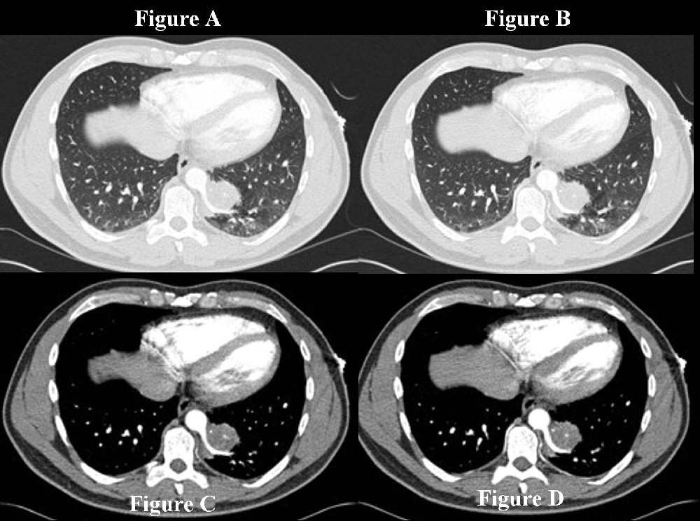
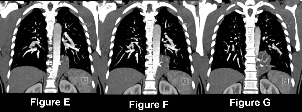
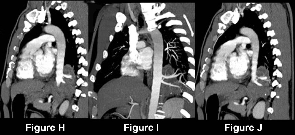
Diagnosis: Intralobar pulmonary sequestration
Differential Diagnosis
- Pneumonia
- Lung abscess
- Bronchiectasis (with secondary infection)
- Infected bulla
Discussion
Background
Pulmonary sequestration refers to lung parenchyma that does not communicate appropriately with the tracheobronchial tree and has its own systemic arterial blood supply. Intralobar sequestrations (ILS) are four times (4X) more common than extralobar sequestration, and occur almost exclusively within the lower lobes, left more often than right. Extralobar sequestrations (ELS) are accessory pulmonary lobes resulting from abnormal foregut budding located outside the normal lung. ELS may occur in the thorax, diaphragm, or abdomen. As opposed to ILS, ELS represent true congenital anomalies and are typically supplied and drained by the systemic circulation.
Etiology
Most ILS represent acquired lesions resulting from lower lobe bronchial obstruction and subsequent distal infection. The concomitant inflammatory process obliterates the normal pulmonary arterial supply to the lesions. As a result, the normal pulmonary ligament arteries (which arise from the descending thoracic aorta) are parasitized to provide a systemic arterial blood supply. Rarely, ILS may receive its arterial blood supply from other sources such as the superior mesenteric artery or even the coronary circulation. A small number of ILS probably do represent true congenital lesions.
Clinical Findings
Patients with ILS are often older children and adolescents. However, approximately 50% of patients with ILS are over the age of 20 years when they first come to clinical presentation, as in this case. Men and women are equally affected. Symptoms associated with ILS may include: recurrent pneumonia, chest pain, chronic cough, sputum production, bronchospasm, and hemoptysis. Patients can also present with acute, chronic, or recurrent lower lobe infection. Approximately 15% of patients with ILS are asymptomatic and diagnosed incidentally.
Pathology
Gross
- Lower lobe (98% of cases); more frequent on left side.
- Thickened overlying visceral pleura; may form adhesions with adjacent structures.
- Densely consolidated lung with internal fluid-filled cysts; cysts may also be filled with mucinous or purulent material.
- Systemic arterial supply; typically coursing within the pulmonary ligament.
- Normal pulmonary venous drainage (95% cases).
Microscopic
- Acute and chronic inflammation, bronchopneumonia, bronchiectasis, fibrosis, and cystic changes.
- Lesion edges contiguous with nonsequestered adjacent lung; sequestered lung may sharply abut normal lung or blend with it diffusely.
- Anomalous feeding arteries.
Imaging Findings
Radiography
- Lower lobe consolidation; most often posterior basal segment; left side more frequently involved than right.
- Consolidation or mass-like region of consolidation may contain air-fluid levels or multi-locular cysts.
- Consolidation or mass-like region of consolidation may have irregular margins or exhibit well-defined borders.
- Surrounding lung parenchyma may be hyperlucent.
- May be associated with impacted bronchi in the vicinity.
- May produce mass effect on neighboring anatomic structures.
CT
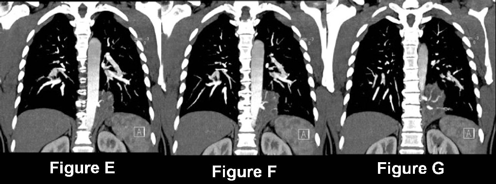
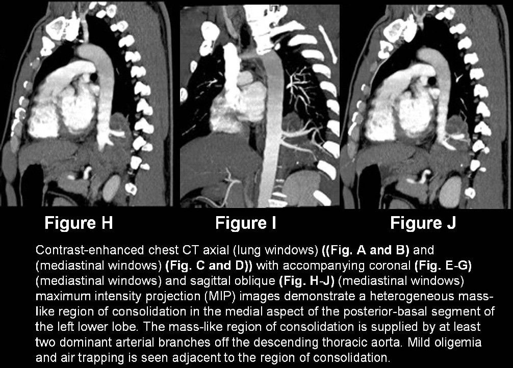
- Heterogeneous or homogenous mass-like region of consolidation medial aspect posterior basal segment lower lobe (Fig. A-J).
- Mass-like consolidation may manifest as soft-tissue alone (Fig. A-J) or as soft-tissue with air and or air-fluid levels, or as a predominately cystic or multi-cystic lesion.
- Smooth, lobular, or irregular borders with the adjacent lung parenchyma (Fig. A-B).
- Surrounding lung parenchyma may be hyperlucent (Fig. A-B).
- Anomalous systemic arterial blood supply, usually from the descending thoracic aorta as one dominant or in some cases, multiple vessels (Fig. C-J); often best demonstrated on multi-planar MIP (Fig. E-J) and 3-D images.
MRI
- Homogenous or heterogeneous mass-like region of consolidation lower lobe.
- May exhibit cystic or multi-cystic foci.
- GRE /MRA sequences may demonstrate the anomalous systemic arterial blood supply.
Treatment
- Pre-operative embolization or ligation of all anomalous feeding vessels.
- Surgical excision of affected lobe (VATS or open thoracotomy)
Prognosis
Caveats
- ILS should be in the differential diagnosis of any patient presenting with recurrent lower lobe infection or a chronic lower lobe radiologic abnormality.
- ILS usually manifests in children and adults with signs and symptoms of infection.
- ELS usually affects neonates and infants who present with respiratory distress.
- ILS is always located inside the lung and may exhibit irregular borders of contact with the normal non-sequestered lung.
- ELS is located outside the lung, does not contain air, and is well-defined.
Suggested Readings
- Choudry R, Salvatore M, Hurie J, Grewal H. Preoperative magnetic resonance imaging for intralobar pulmonary sequestration. Pediatr Surg. 2006;41(4):872-874.
- Deguchi E, Furukawa T, Ono S, Aoi S, Kimura O, Iwai N. Intralobar pulmonary sequestration diagnosed by MR angiography. Pediatr Surg Int 2005; 21(7): 576-577.
- Ferretti GR, Jouvan FB, Coulomb M. MDCT demonstration of intralobar pulmonary sequestration of the right upper lobe in an adult. AJR 2005; 185(6): 1663-1664.
- Frazier AA, Rosado-de-Christenson ML, Stocker TJ, Templeton PA. Intralobar sequestration: radiologic-pathologic correlation. RadioGraphics 1997; 17: 725-745.
- Parker MS, Abbott GF. Developmental Anomalies: Tracheobronchial Anomalies. In: Teaching Atlas of Chest Imaging. New York: Thieme Medical Publishers, Inc. 2006; 33-47.
- Tsitouridis I, Tsinoglou K, Cheva A, Papapostolou P, Efthimiou D, Moschialos L. Intralobar pulmonary sequestration with arterial supply from the coronary circulation. J Thorac Imaging. 2005; 20(4):313-315.
Original case written by its authors at Virginia Commonwealth University and published at this address as part of a weekly teaching collection. Reproduced here as an archive.