Diagnosis: Type III ductus diverticulum
Differential Diagnosis
None
Discussion
Background
The increased use of MDCT has led to the recognition of numerous vascular variants that may mimic acute aortic injuries. The most common variants include: type ductus diverticulum; atypical ductus diverticulum; aortic spindle; and branch vessel infundibula.
Ductus diverticulum is either a remnant of the ductus arteriosum or the right dorsal aortic root. The most common diagnostic challenge for radiologists on trauma chest CT’s is the differentiation of a post-traumatic aortic isthmus pseudoaneurysm from a normal type III ductus diverticulum. Both aortic entities occur in roughly the same anatomic location often leading to diagnostic confusion. There are 4 distinct variations in the contour of the aortic isthmus as follows:
Type I - a concave contour.
Type II-mild straightening or convexity without a discrete bulge.
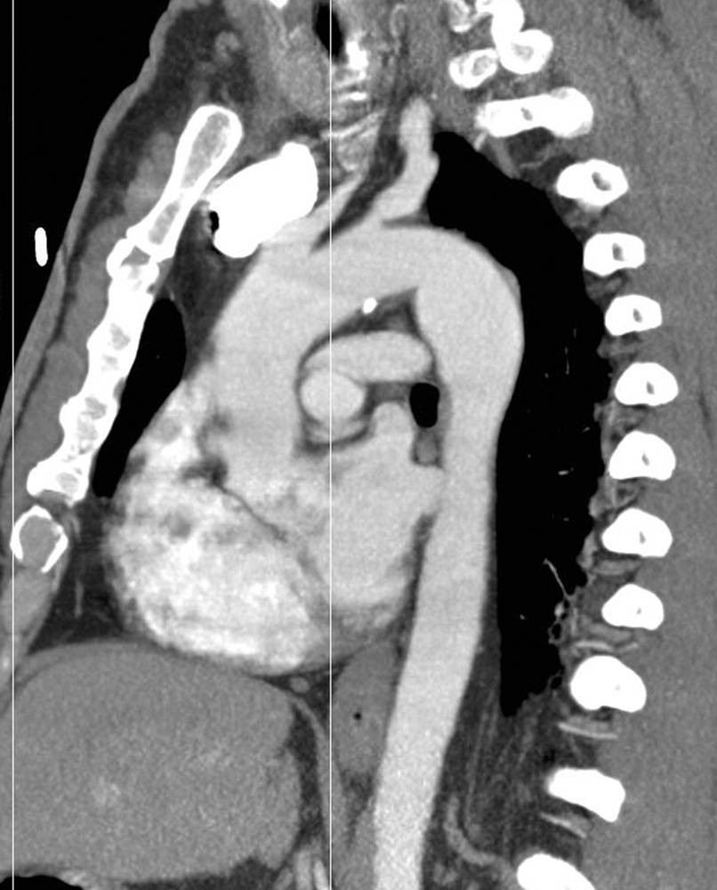
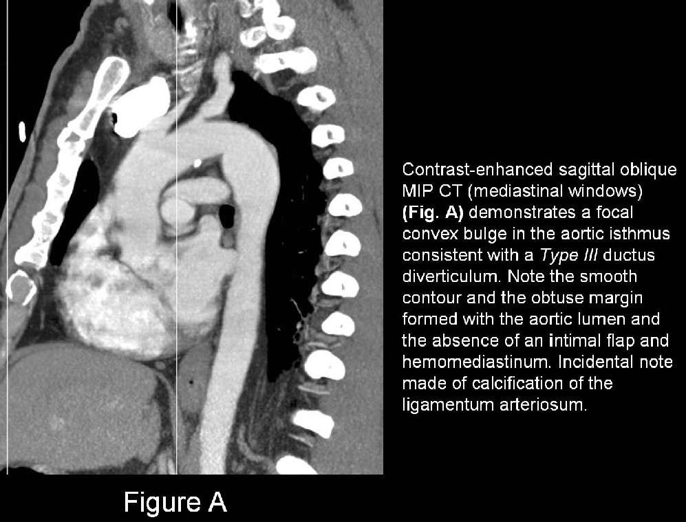
Type III - a discrete focal bulge referred to as the ductus diverticulum (Fig. A)
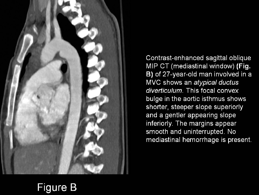
Atypical ductus diverticulum- often causes even more diagnostic confusion; characterized superiorly by a shorter, steeper slope and inferiorly by a more typical, gentler slope (Fig. B).
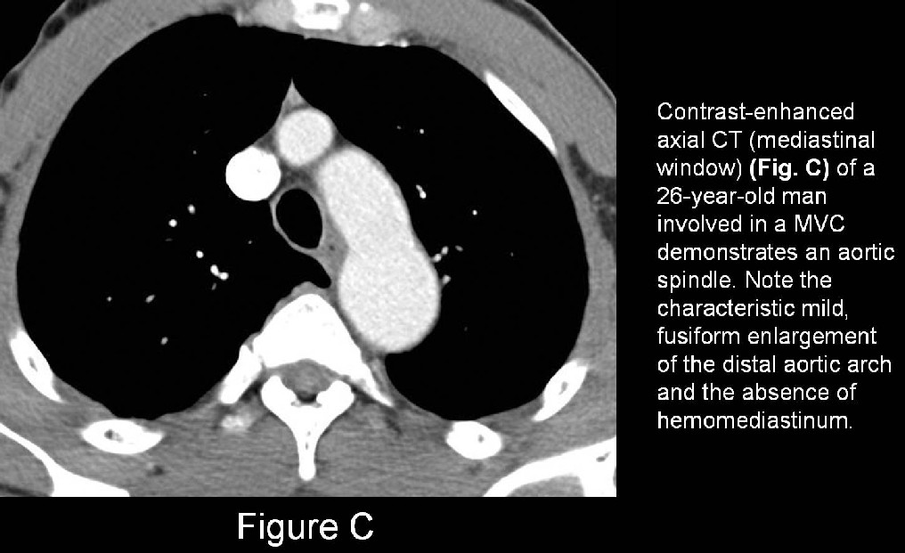
Aortic spindle manifests as a fusiform dilatation of the aorta immediately distal to the isthmus. That is the region between the left subclavian artery origin and the point of attachment of the ligamentum arteriosum) (Fig. C).
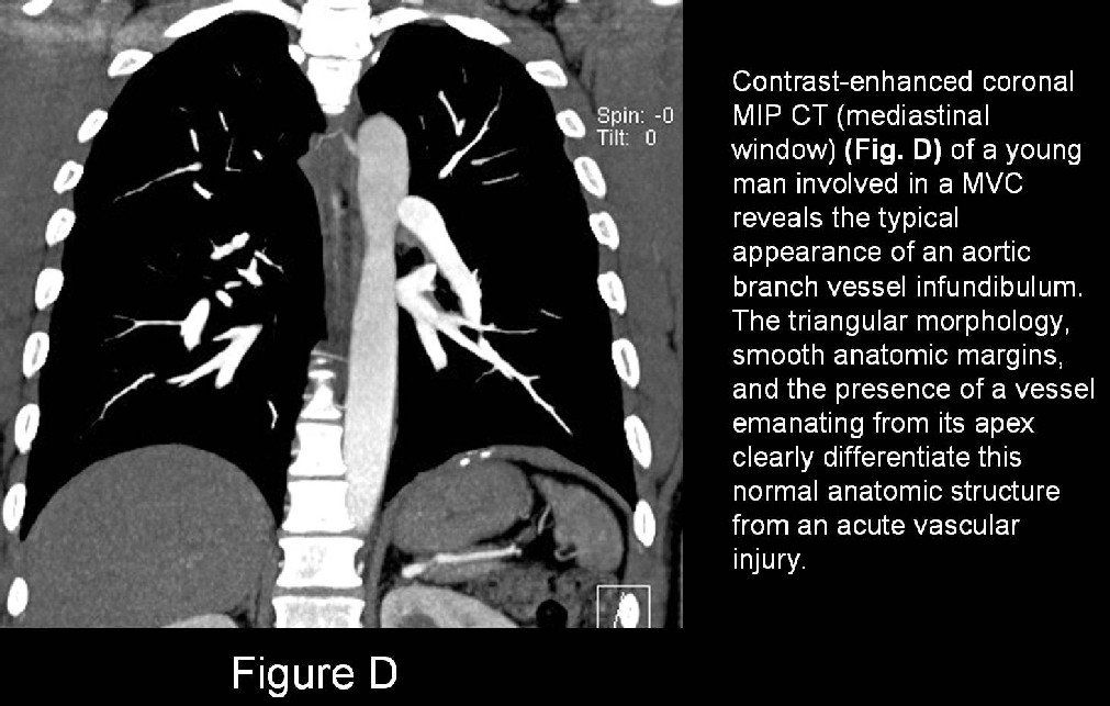
Infundibula of the aortic branch vessels, including the brachiocephalic artery, bronchial artery, intercostal arteries (i.e., most commonly the right third), left common carotid and left subclavian arteries, may simulate acute traumatic injuries or pseudoaneurysms. Infundibula are recognized by their triangular anatomic morphology, smooth margins, and by the presence of a vessel emanating from their apex (Fig. D).
Imaging Features
CT/MR
Type III Ductus Diverticulum
- Best appreciated on sagittal and sagittal oblique MIP CT images (Fig. A).
- Focal convex bulge with a smooth contour that forms an obtuse angle with the aortic lumen (Fig. A).
- Intimal flaps and hemomediastinum are not present (Fig. A).
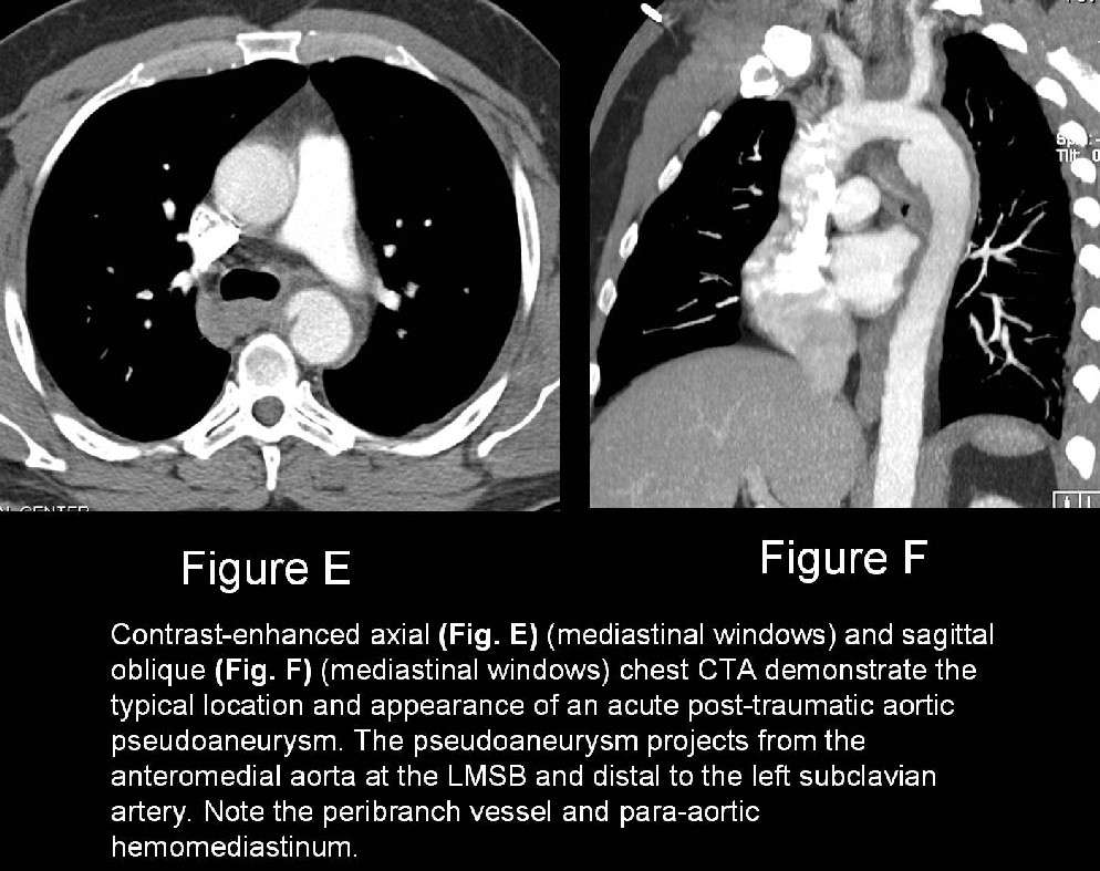
- In contrast, post-traumatic pseudoaneurysm manifests as an irregular out pouching from the aortic lumen, displaying more acute margins. Intimal flaps and mediastinal hemorrhage are usually present (Fig. E; F).
- On axial CT images, ductus diverticulum demonstrates a smooth transition between contiguous slices whereas a post-traumatic pseudoaneurysm is variable in shape with sharp and more irregular margins.
Atypical Ductus Diverticulum
- Shorter, steeper slope superiorly, and a more typical, gentler slope, inferiorly.
- Smooth uninterrupted margins aid in differentiation from true post-traumatic aortic pseudoaneurysm (Fig. B).
Aortic Spindle
- Fusiform dilatation of aorta immediately distal to isthmus (Fig. C).
- Mild, fusiform enlargement of distal aortic arch (Fig. C).
- MPR and MIP images acquired along the vascular axis are helpful supplements; reveal a normal caliber aortic arch and proximal descending thoracic aorta.
Branch Vessel Infundibula
- Recognized by their triangular anatomic morphology (Fig. D).
- Smooth margins (Fig. D).
- Presence of a vessel emanating from their apex (Fig. D).
- MIP and 3-D reformations are useful confirmatory images; delineate origin and course of the vessel related to the infundibulum (Fig. D).
Treatment
- Represent anatomic variations in normal anatomy; no management indicated.
Prognosis
Caveats
- Radiologists must be mindful of normal anatomic variants that may simulate acute aortic injury.
Suggested Readings
- Goodman PC, Jeffrey RB, Minagi H, Federle MP, Thomas AN. The angiographic evaluation of the ductus diverticulum. Cardiovasc Intervent Radiol 1982; 5:1-4.
- Grollman JH. The aortic diverticulum: a remnant of the partially involuted dorsal aortic root. Cardiovasc Intervent Radiol 1989; 12:14-17.
- Macura KJ, Corl FM, Fishman EK, Bluemke DA. Pictorial Essay: Pathogenesis in acute aortic syndromes: aortic aneurysm leak and rupture and traumatic aortic transection. AJR Am J Roentgenol 2003; 181: 303-307.
Original case written by its authors at Virginia Commonwealth University and published at this address as part of a weekly teaching collection. Reproduced here as an archive.