Diagnosis: Exogenous Lipoid Pneumonia
Differential Diagnosis
- Endogenous lipoid pneumonia; in the setting of proximal or distal airway obstruction
- Pseudolipoid pneumonia; an air-bubble artifact associated with collapse of airspaces.
- Infection
- Primary lung cancer
- Pulmonary lymphoma
- Other fat-containing lesions
Differential Diagnosis other Intrathoracic Fat-containing Lesions:
- Hamartoma
- Lipoma
- Liposarcoma
- Germ cell tumors
- Mediastinal lipomatosis
- Thymolipoma
- Diaphragmatic hernias, fat herniation
Discussion
Background
Exogenous lipid pneumonia (ELP) is an uncommon cause of parenchymal consolidation secondary to the chronic aspiration or inhalation of animal, vegetable, or petroleum-based oils or fats.
Etiology
Mineral oil is the most common irritant and has the ability to inhibit the cough reflex and ciliary motility resulting in a “silent” inhalation. ELP is more common in the elderly, young children, and debilitated patients. Two-thirds of patients have a predisposition to aspiration from swallowing dysfunction, structural esophageal abnormalities, or altered consciousness. Infants with feeding difficulties may aspirate oil used as a lubricant. Elderly patients with constipation may aspirate oil ingested as a laxative. Other potential sources include: oil-based nose drops, camphor and eucalyptus oil-based cough suppressants and nasal decongestants, lip gloss, and petroleum jelly applied to the face and nose for various reasons. Aspiration of animal oils may be associated with the ingestion of milk or milk products, and cod liver and shark liver oil. Most vegetable oil aspirations occur while eating or with regurgitation of gastric contents. Pure vegetable oil aspiration is uncommon. An unusual form of ELP is seen in “fire-eaters” who use liquid paraffin to help generate the fire. The pathogenesis of mineral and vegetable oil-related fibrosis is unknown. The release of lysosomal enzymes by lipid-laden macrophages may be responsible. Animal fats are hydrolyzed by lung lipases into fatty acids that may cause acute hemorrhagic pneumonitis.
Clinical Findings
Most patients with mineral oil-related ELP are asymptomatic and present as an incidental radiologic finding. The most frequent signs and symptoms associated with animal and vegetable oil-related ELP mirror those of acute bronchopneumonia and include: fever (39%); weight loss (34%); cough (64%); dyspnea (50%); and crepitations (45%). Lung function is usually normal or shows a restrictive pattern. DLCO is frequently diminished. BAL fluid often shows non-specific increases in lymphocytes, eosinophils, multinucleated giant cells containing lipid droplets, and large numbers of lipid-laden macrophages. This latter finding is highly suggestive of chronic aspiration or lipoid pneumonia.
Pathology
Acute: alveoli fill with lipid
Subacute:
1 week: Lipids phagocytosed by macrophages and other inflammatory cells.
>1week: Distention of alveolar walls can lead to acute and chronic pneumonitis. Clearance occurs through lymphatics and mucociliary pathways.
Chronic:
Weeks to months: Macrophages transport oil from alveoli to interlobular septa leading to granuloma formation and pulmonary fibrosis.
Imaging Findings
Radiography
- Variable patterns depending on the volume and nature of aspirate
- Preferential involvement of dependent lung zones
- Cavitation in cases of secondary anaerobic infection
- Acute aspiration of large amounts of animal oil:
- Extensive ground-glass opacities and/or
- Frank regions of consolidation
- Aspiration of mineral oil:
- Ground-glass opacities
- Septal lines
- Lung mass resembling primary lung cancer
- Aspiration of vegetable oil:
- Inconspicuous
- Atelectasis
- Variable sized poorly defined nodular opacities
CT/HRCT
- Preferential involvement of dependent lung zones
- Ground-glass opacities
- Initial pattern (50%) with centrilobular or panlobular distribution due to aspiration
- Ground-glass and interstitial opacities forming a “crazy-paving pattern” (33%)
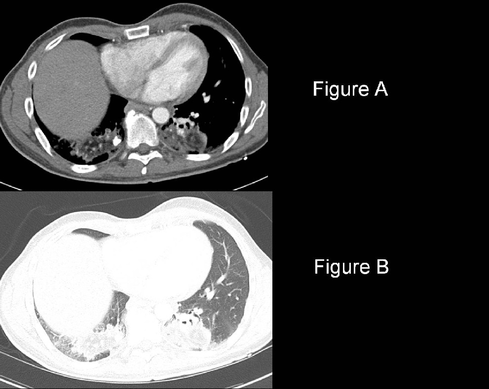
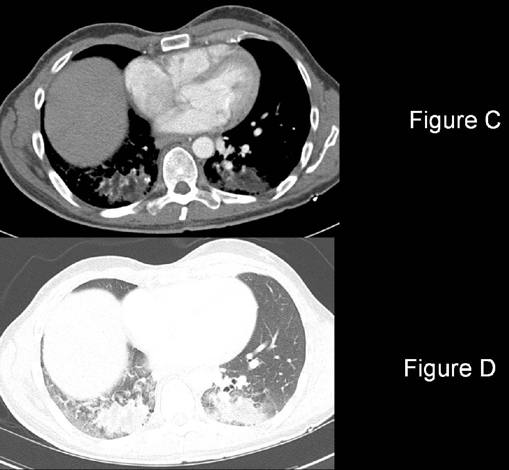
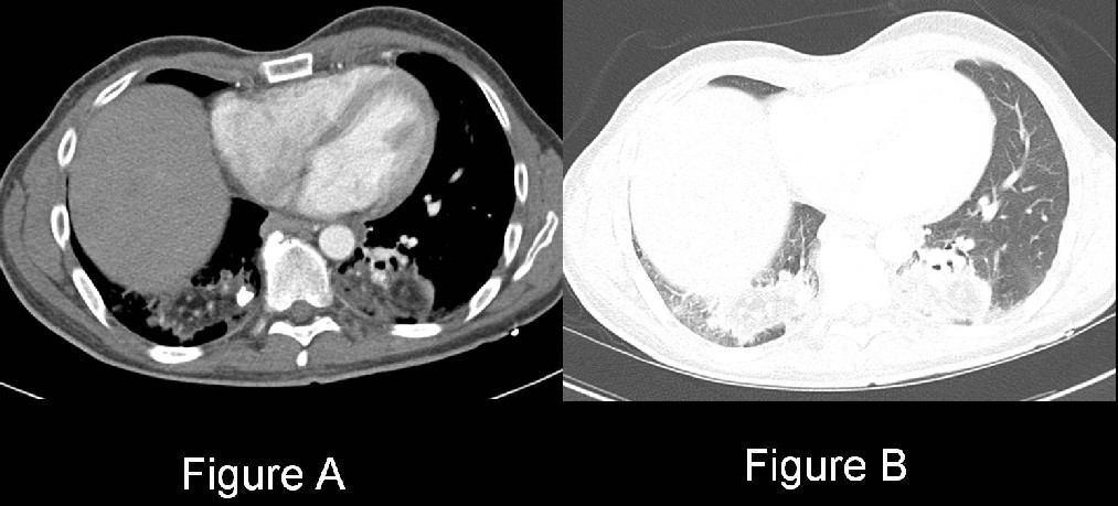
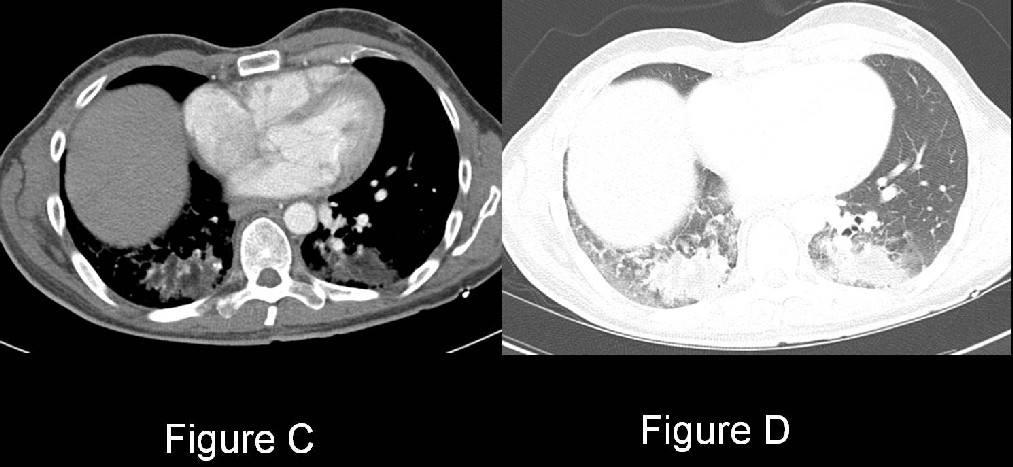
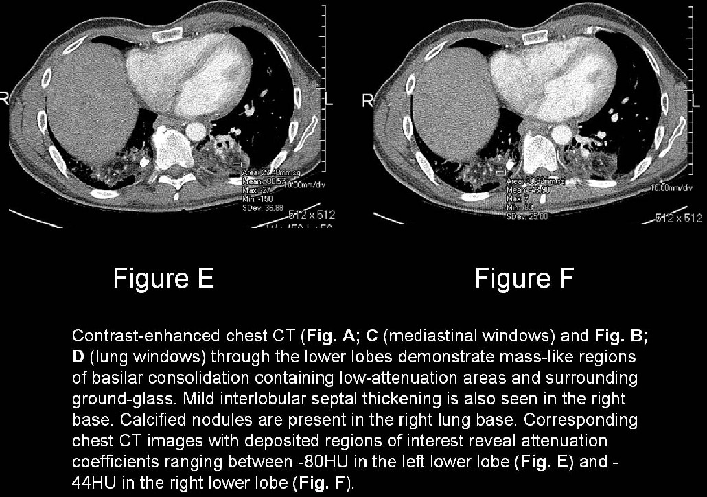
- Patchy unilateral or bilateral airspace consolidation(s) (90%) (Fig. A-D)
- Consolidation with internal low or fat attenuation (-10 to -150 HU) (i.e., attenuation less than that of the chest wall musculature but often greater than that of subcutaneous fat) (80%) (Fig. A; C: E; F)
- Centrilobular nodules
- Foci of calcification; rare (Fig. A; C: E; F)
- Chronic cases: juxtapleural pulmonary fibrosis; architectural distortion; honeycombing; volume loss
MR
- High signal intensity on both T1- and T2-weighted images (i.e., lipid content)
- Chemical-shift/in-phase/out-of-phase imaging useful to make a specific diagnosis
PET
- FDG-PET scans reveal standard uptake values (SUV) suggestive of malignancy
Management
- Mainstay: removal of the offending agent.
- Correction of underlying defects or abnormalities that predispose to aspiration.
- Oral steroids; helpful in select cases
- Prednisone and whole lung lavage; may be successful in cases of diffuse ELP
Prognosis
- ELP may be complicated by superimposed infection, which should be suspected when cavitary nodules are present.
- Increased risk of infection with nontuberculous mycobacteria (e.g., Mycobacterium fortuitum, Mycobacterium chelonae) and of developing primary lung cancer.
- Clinical course and outcome of animal and vegetable oil aspiration: similar to that of aspiration of gastric contents.
CAVEATS
- ELP predominantly affects the lower lobes; posterior segments of the upper lobes; superior segments of lower lobes; and middle lobes are affected less often.
- ELP may mimic primary and secondary lung neoplasia; the appropriate diagnosis is dependent upon recognition of fat or low-attenuation areas in the consolidation.
- Increased risk of infection with Mycobacterium fortuitum and Mycobacterium chelonae and of developing primary lung cancer.
- ELP may be a cause of false-positive F-18-fluorodeoxyglucose (FDG) uptake on positron emission tomography (PET) imaging.
Suggested Readings
- Fox BD, Shechtman I, Shitrit D, Bendayan D, Kramer MR. A “fat chance” its malignant: lipoid pneumonia simulating lung cancer on PET scan. Thorax 2007; 62(5):464.
- Gaerte SC, Meyer CA, Winer-Muram HT, Tarver RD, Conces DJ Jr. Fat containing lesions of the chest. RadioGraphics 2002; 22(S): S61-78.
- Kitchen JM, O'Brien DE, McLaughlin AM. Perils of fire eating. An acute form of lipoid pneumonia or fire eater’s lung. Thorax 2008; 63(5):401, 439.
- Mokhlesi B, Angulo-Zereceda D, Yaqhmai V. False-positive FDG-PET scan secondary to lipoid pneumonia mimicking a solid pulmonary nodule. Ann Nucl Med 2007; 21(7):411-4. Epub 2007 Sep 25.
- Parker MS, Rosado-de-Christenson ML, Abbott GF. Exogenous Lipoid Pneumonia. In: Teaching Atlas of Chest Imaging. New York, NY: Thieme Medical Publishers, Inc., 2006: 481-484.
- Travis WD, Colby TV, Koss MN, Rosado-de-Christenson ML, Muller NL, King TE Jr. Diffuse parenchymal lung diseases. In: King DW, ed. Atlas of Nontumor Pathology: Non-Neoplastic Disorders of the Lower Respiratory Tract, fascicle 2, series 1. Washington, DC: American Registry of Pathology and Armed Forces of Pathology: 187-196.
- Zanetti G, Marchiori E, Gasparetto TD, Escuissato DL, Soares Souza A Jr. Lipoid pneumonia in children following aspiration of mineral oil used in the treatment of constipation: high-resolution CT findings in 17 patients. Pediatr Radiol 2007; 37(11):1135-9. Epub 2007 Sep 18.
Original case written by its authors at Virginia Commonwealth University and published at this address as part of a weekly teaching collection. Reproduced here as an archive.