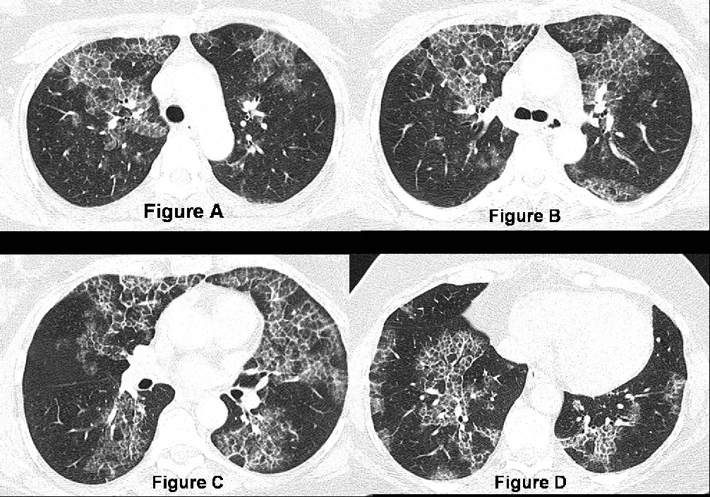
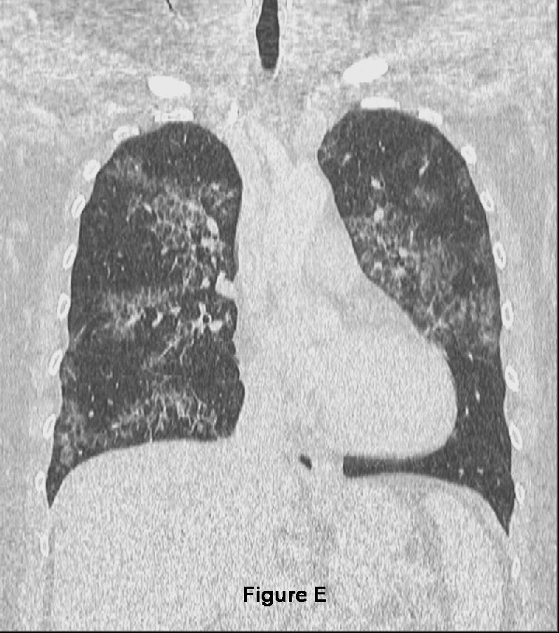
Unenhanced chest CT axial (Figures A-D; lung windows) and coronal (Figure E) images demonstrate bilateral symmetric ground-glass and smooth reticular opacities diffusely throughout the lungs without a zonal predilection. The involved regions of lung parenchyma are sharply demarcated from the adjacent normal lung. This combination of ground-glass with concomitant reticular opacities creates a pattern of disease called “crazy-paving”. Note the preservation of lung volume and the absence of lymphadenopathy and pleural effusion. Diagnosis: Pulmonary Alveolar Proteinosis
Differential Diagnosis
- Edema
- Cardiogenic
- ARDS
- Acute Interstitial Pneumonia (AIP)
- Infection
- Pneumocystis jiroveci pneumonia
- Viral pneumonia
- Mycoplasma pneumonia
- Bacterial pneumonia
- Organizing Pneumonia
- Cryptogenic Organizing Pneumonia (COP) / Bronchiolitis Obliterans Organizing Pneumonia (BOOP)
- Neoplasia
- Bronchioloalveolar carcinoma (BAC)
- Other
- Hemorrhage
- Pulmonary Alveolar Proteinosis
- Sarcoidosis
- Non-specific Interstitial Pneumonia (NSIP)
- Lipoid pneumonia
- Subacute radiation therapy-related pneumonitis (XRT)
Discussion
Background
Pulmonary Alveolar Proteinosis (PAP), also known as alveolar lipoproteinosis and alveolar phospholipoproteinosis, is a rare lung disorder (estimated prevalence of 1 case per 100,000 population) characterized by the abnormal accumulation of lipid-rich granular eosinophilic material within the alveoli.
Etiology
The etiology of PAP is unknown. However, three forms are recognized. These include primary (idiopathic), secondary, and congenital varieties. Primary PAP is the most common form, accounting for 90% of cases, and occurs in isolation. Secondary PAP (5%-10%) occurs with the industrial inhalation of mineral dusts (e.g., silica, titanium oxide, aluminum) and insecticides; various hematologic malignancies, and immunodeficiency syndromes including AIDS. An association with cigarette smoking has also been suggested. Congenital PAP is rare (2%). Affected neonates are deficient in surfactant-associated protein B (SP-B).
Clinical Findings
Most patients presenting with PAP are 20-50-years old and have a gradual onset of symptoms including a persistent dry cough, progressive dyspnea, fatigue, malaise, weight loss, low-grade fever and or night sweats. Expectoration of gelatinous material has been reported. However, up to 30% of affected patients are asymptomatic, even in the setting of profoundly abnormal chest radiography. Physical exam findings may include fine end-inspiratory crackles. Pulmonary function tests (PFT’s) often reveal impaired diffusion and mild to moderate restrictive physiology. PAP is 4X more common in males than females.
Pathology
Gross
- Heavy, congested lungs.
- Nodular consolidations of varying sizes.
Microscopic
- Patchy or diffuse accumulation of eosinophilic periodic acid-Schiff (PAS)-positive proteinaceous material with alveolar spaces.
- Mild interstitial thickening but no fibrosis.
Imaging Findings
Radiography
- Bilateral, symmetric, patchy and diffuse ground-glass opacities and air-space consolidations.
- Nodular or reticular opacities.
- Lower lobe predilection.
- Relative sparing of costophrenic angles and apices.
- Preserved lung volumes.
- Lymphadenopathy and pleural effusion; rare.
CT / HRCT
- Diffuse patchy bilateral ground-glass with superimposed smooth reticular opacities or septal thickening (“crazy-paving” pattern) (Figure A-E).
- Areas of affected lung are sharply demarcated from adjacent uninvolved or normal lung (Figure A-E).
- Multifocal nodules or confluent areas of consolidation may occur with concomitant superimposed infection.
Management
- Depends on the degree of physiological impairment, underlying concomitant diseases or associated factors and the presence of coexisting infection(s).
- Indications for mechanical removal of the lipoproteinaceous-rich material from the alveoli by bilateral, sequential whole lung lavage with isotonic NaCl solution and/or repeated lobar lavage via bronchoscopy include:
- Alveolar-arterial oxygen gradient ≥40 mm Hg
- PaO 2 <65 mm Hg
- Dyspnea and hypoxemia at rest or with exercise
- Lung transplantation is reserved for:
- Adult patients with end-stage interstitial fibrosis and complicating cor pulmonale
- Congenital PAP
Prognosis
- Overall prognosis for primary PAP is very good.
- Often dramatic clinical improvement and or remission with one-time whole-lung lavage.
- Relapses may necessitate repeat lavage(s).
- These patients often have a poorer outcome complicated by
- Interstitial fibrosis
- Respiratory failure
- Cor pulmonale
- Complicating lung infections include:
- N. asteroides
- M. tuberculosis
- Mycobacterium avium-intracellulare
- S. pneumoniae
- Aspergillus sp.
- Pneumocystis jiroveci
- Candida
- C. neoformans
- H. capsulatum
- Cytomegalovirus
Caveats
- Primary PAP may occur in isolation.
- 30% patients are asymptomatic, even in the setting of profoundly abnormal chest radiography.
- Whole lung lavage is the treatment of choice in primary PAP.
- The “crazy-paving” pattern is not pathognomonic of PAP but has a broad differential diagnosis which requires clinical and laboratory correlation to appropriately narrow.
References
- Frazier AA, Franks TJ, Cooke EO, Mohammed TH, Pugatch RD, Galvin JR. From the archives of AFIP: Pulmonary alveolar proteinosis. RadioGraphics 2008; 28:883-899.
- Holbert JM, Costello P, Li W, Hoffman RM, Rogers RM. CT features of pulmonary alveolar proteinosis. AJR 2001; 176(5):1287-1294.
- Rossi SE, Erasmus JJ, Volpacchio M, Franquet T, Castiglioni T, McAdams HP. “Crazy-Paving” pattern at thin-section CT of the lungs: radiologic-pathologic overview. RadioGraphics 2003; 23:1509-1519.
- Parker MS, Rosado-de-Christenson ML, Abbott GF. Diffuse Lung Disease. In: Teaching Atlas of Chest Imaging. New York, NY: Thieme Medical Publishers, Inc., 2006: 455-458.
- Travis WD, Colby TV, Koss MN, Rosado-de-Christenson ML, Müller NL, King TE Jr. Idiopathic interstitial pneumonia and other diffuse parenchymal lung diseases. In: King DW, ed. Atlas of Nontumor Pathology: Non-Neoplastic Disorders of the Lower Respiratory Tract, fascicle 2, series 1. Washington, DC: American Registry of Pathology and Armed Forces Institute of Pathology: 49-231
Original case written by its authors at Virginia Commonwealth University and published at this address as part of a weekly teaching collection. Reproduced here as an archive.