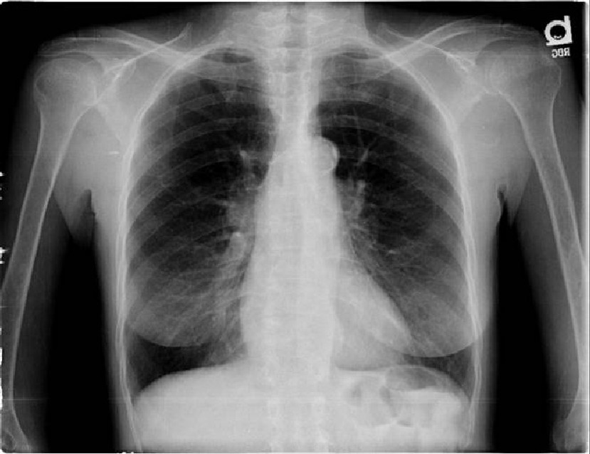
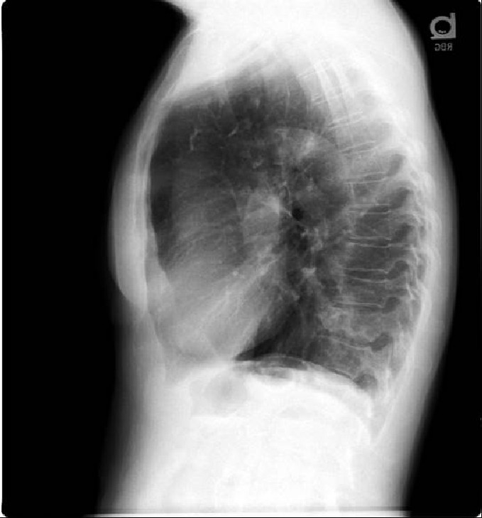
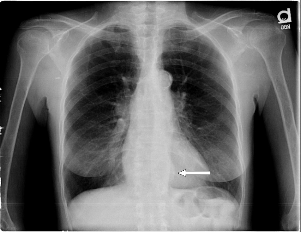
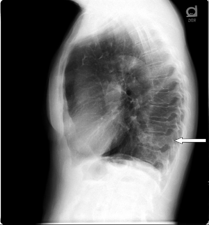
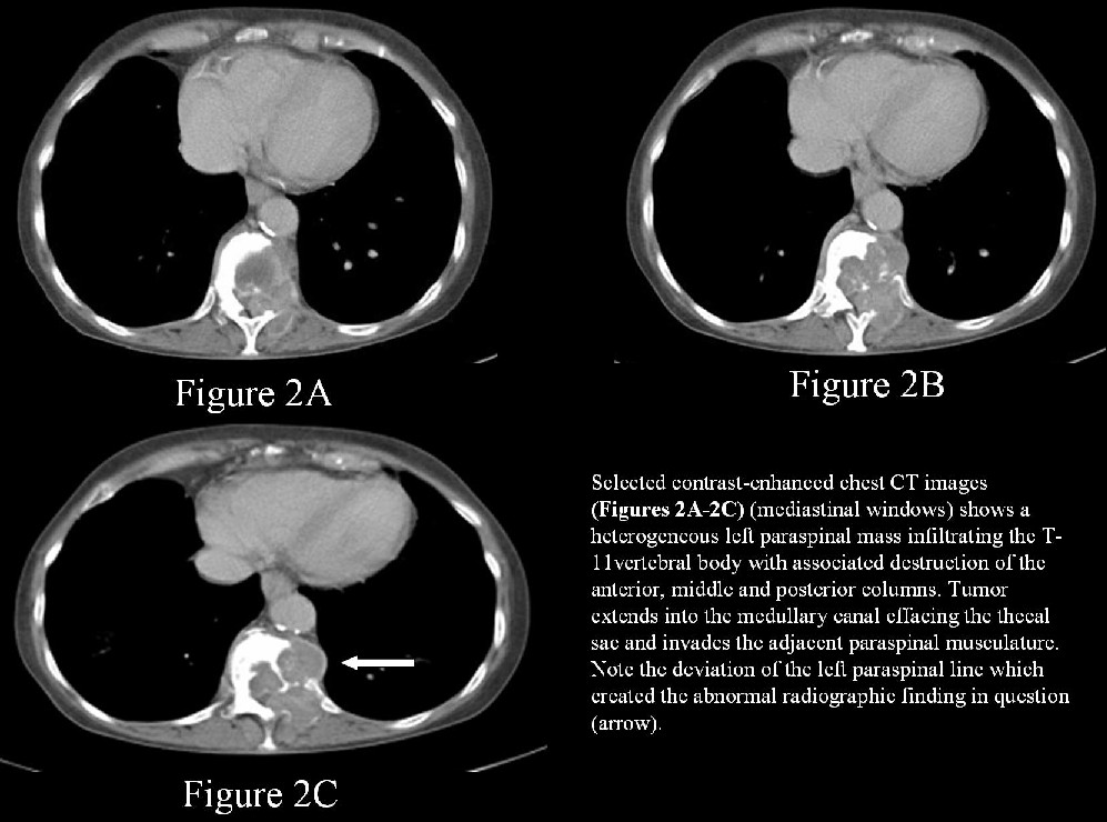
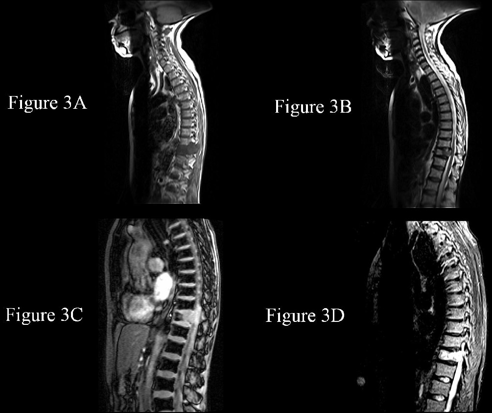
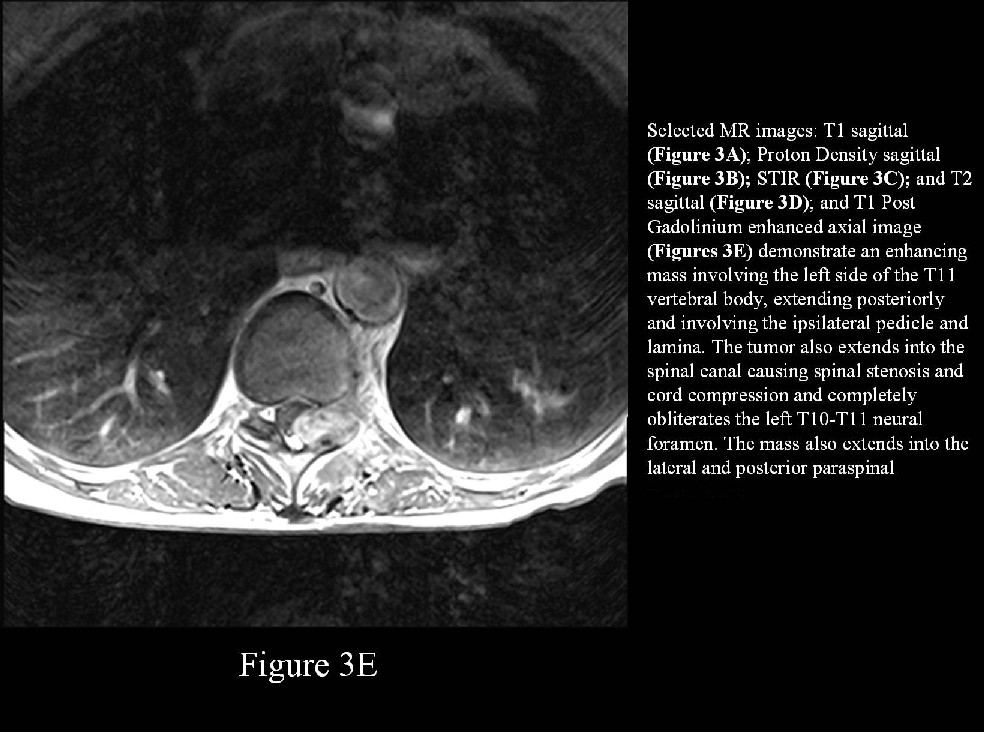
Left paraspinal mass-metastatic carcinosarcoma to the thoracic spine (biopsy proven); unknown primary.
PA and lateral chest radiography demonstrates a focal convex bulge in the left lateral paraspinal line with associated loss of height and destruction of the T-11 vertebral body (arrows). There is a background of underlying obstructive lung disease and atheromatous changes are present in the thoracic aorta and its branch vessels. Diagnosis: Left paraspinal mass-metastatic carcinosarcoma to the thoracic spine (biopsy proven); unknown primary.
Differential Diagnosis
Benign Lesions
- Osteophytes
- Mediastinal lipomatosis
- Tortuosity of the descending thoracic aorta
- Mediastinal hematoma
- Extramedullary hematopoesis
- Uphill paraesophageal varices
- Posterior mediastinal or paraspinal fluid collections
- Mediastinal extension of pancreatic pseudocyst
- Descending thoracic aortic aneurysms
- Azygos and hemiazygos vein dilation
- Vertebral body fractures with associated hematoma
- Infectious spondylitis / Pott’s disease
Paraspinal mass lesions
- Neurenteric cyst
- Thoracic lateral meningocele
- Neurofibroma
- Neurilemmoma
- Ganglioneuroma
- Neuroblastoma
- Pheochromocytoma
- Chemodectoma
Metastasis to thoracic spine from the following primary lesions
- Lung - 31%
- Breast - 24%
- GI tract - 9%
- Prostate - 8%
- Lymphoma - 6%
- Melanoma - 4%
- Unknown - 2%
- Kidney - 1%
Background
The configurations and interrelationships of the anatomic structures in the lung, mediastinum, and pleura forms the basis of identifying lines and stripes on chest radiography, which plays a valuable role in establishing a diagnosis before proceeding to cross-sectional imaging studies such as CT. The inability to recognize radiographic abnormalities resulting from displacement of one of these lines or stripes may lead to failure to request a potentially valuable CT examination. It is important to be familiar with the anatomic basis of these mediastinal lines and stripes and be able to recognize their normal and as well abnormal appearances and the differential diagnosis associated with such.
Discussion
What is the paraspinal line?
Right paraspinal line -The right paraspinal line is formed by the right lung and pleura coming in tangential contact with the posterior mediastinal soft tissues. The right paraspinal line appears straight and typically extends from approximately T-8-T-12.The right paraspinal line can be identified on up to 23% of normal frontal radiographs.
Left paraspinal line - The left paraspinal line is formed by tangential contact of the left lung and pleura with the posterior mediastinal fat, left paraspinal muscles, and adjacent soft tissues. The left paraspinal line extends vertically from the aortic arch to the diaphragm and typically lies medial to the lateral wall of the descending thoracic aorta. The left paraspinal can be seen on up to 41% of normal frontal radiographs. It’s more frequent conspicuity is due to the presence of the descending thoracic aorta on the left, which accentuates the tangential contact of the left lung necessary to produce the lung-mediastinum interface.
What comprises the paraspinal line?
Right paraspinal line -The right paraspinal line is not a true mediastinal line. It actually represents an interface between the right lung and the posterior mediastinal fat and soft tissues. A thin white line enhancing the edge of the right paraspinal line does not really exist but is perceived by the observer optically and is a positive Mach band phenomenon.
Left paraspinal line - The left paraspinal line does indeed represent a true lung-mediastinum Interface but likewise is associated with a positive Mach band phenomenon, having the appearance of a line etched in white.
Clinical Findings
Carcinosarcoma of the bladder
- Gross, painless hematuria.
Carcinosarcoma of the uterus
- Majority (80%) of patients present with abnormal postmenopausal uterine bleeding.
- May present as a rapidly enlarging uterine mass.
Carcinosarcoma of the lung
- Cough
- Hemoptysis
- Weight loss
- Constitutional symptoms
Pathology
Carcinosarcoma of the bladder
- Rare tumors.
- Usually occurring in middle-aged men.
- Highly malignant containing malignant mesenchymal and epithelial elements.
- Mesenchymal elements are usually
- Chondrosarcoma
- Osteosarcoma
- Epithelial elements may be
- Transitional cell carcinoma
- Squamous cell carcinoma
- Adenocarcinoma
Carcinosarcoma of the uterus (Malignant Mixed Müllerian Tumor)
- Represent about 50% of all uterine sarcomas, or 3% of all uterine neoplasms.
- Occur at a median age of 66 years.
- Contain mixture of epithelial and mesenchymal elements.
- Epithelial elements are typically adenocarcinoma.
- Mesenchymal component may be classified
- Homologous
- Endometrial stromal sarcoma
- Fibrosarcoma
- Leiomyosarcoma
- Undifferentiated sarcoma
- Heterologous
Carcinosarcoma of the lung
- Extremely rare.
- Composed of mixture of epithelial and mesenchymal neoplastic cells.
- Contains malignant mature epithelium (non-small cell carcinoma) surrounded by malignant-appearing mesenchymal elements, either primitive or differentiated sarcoma.
- Reported mostly in smokers and in middle-aged and older men
- Behaves like bronchogenic carcinoma.
- May present as endobronchial or parenchymal mass lesion.
- Metastases to lymph nodes, bones, brain, lung, and adrenals as well as local invasion are common.
Treatment
Carcinosarcoma of the uterus
- Surgical resection.
- Adjuvant radiation therapy reserved for patients at risk for pelvic recurrence.
- Radiation therapy for early-stage endometrial carcinoma shown to decrease the rate of local recurrence.
- Definitive radiation reserved for patients who are medically inoperable or who refuse surgical resection.
Carcinosarcoma of the bladder
- Cystectomy
- Radiation and/or chemotherapy
Carcinosarcoma of the lung
- Surgical resection
- Palliation with radiation therapy for patients unsuited for surgery
- Chemotherapy has a limited role
- Palliation of metastatic disease with radiation therapy or chemotherapy
Treatment for vertebral metastases
- Corpectomy
- Methymethacrylate injection
- Spinal fusion for stabilization
Prognosis
- Poor prognosis even with aggressive treatment
Caveats
- Even with the increased dependence on CT in the evaluation of chest disease, traditional chest radiography remains a valuable tool.
- Radiologists must be familiar with the anatomic basis of the mediastinal lines and stripes seen on chest radiography.
- Recognition of the normal and abnormal appearances of these lines and stripes helps to develop an appropriate differential diagnosis prior to obtaining additional information with cross-sectional imaging.
Suggested Readings
- Lines and Stripes: Where Did They Go? —From Conventional Radiography to CT. Jerry M. Gibbs, MD, Chitra A. Chandrasekhar, MBBS, Emma C. Ferguson, MD, Sandra A. A. Oldham, MD. RadioGraphics 2007; 27:33–48 ? Published online 10.1148/rg.271065073
- Mason R J, Murray J F. Mason: Murray & Nadel's Textbook of Respiratory Medicine, 4th Ed. Chapter 45 - Lymphoma, Lymphoproliferative Diseases, and Other Primary Malignant Tumors. Saunders Elsevier, 2005.
- Abeloff MD, Armitage JO, Niederhuber JE. Abeloff's Clinical Oncology, 4th ed. Chapter 92 – Cancer of the Endometrium. Churchill Livingstone Elsevier, 2004.
- WeinA J, Kavoussi L R; Campbell-Walsh Urology, 9th Ed.; Chapter 75 Urothelial Tumors of the Bladder- Nonurothelial tumors of the bladder. Saunders Elsevier, 2007.
- Webb RW, Brant W, Major N, Fundamental of Body CT, 3rd Ed.; Chapter 4 - Mediastinum: Lymph node abnormalities and masses. Saunders, 2005.
Original case written by its authors at Virginia Commonwealth University and published at this address as part of a weekly teaching collection. Reproduced here as an archive.