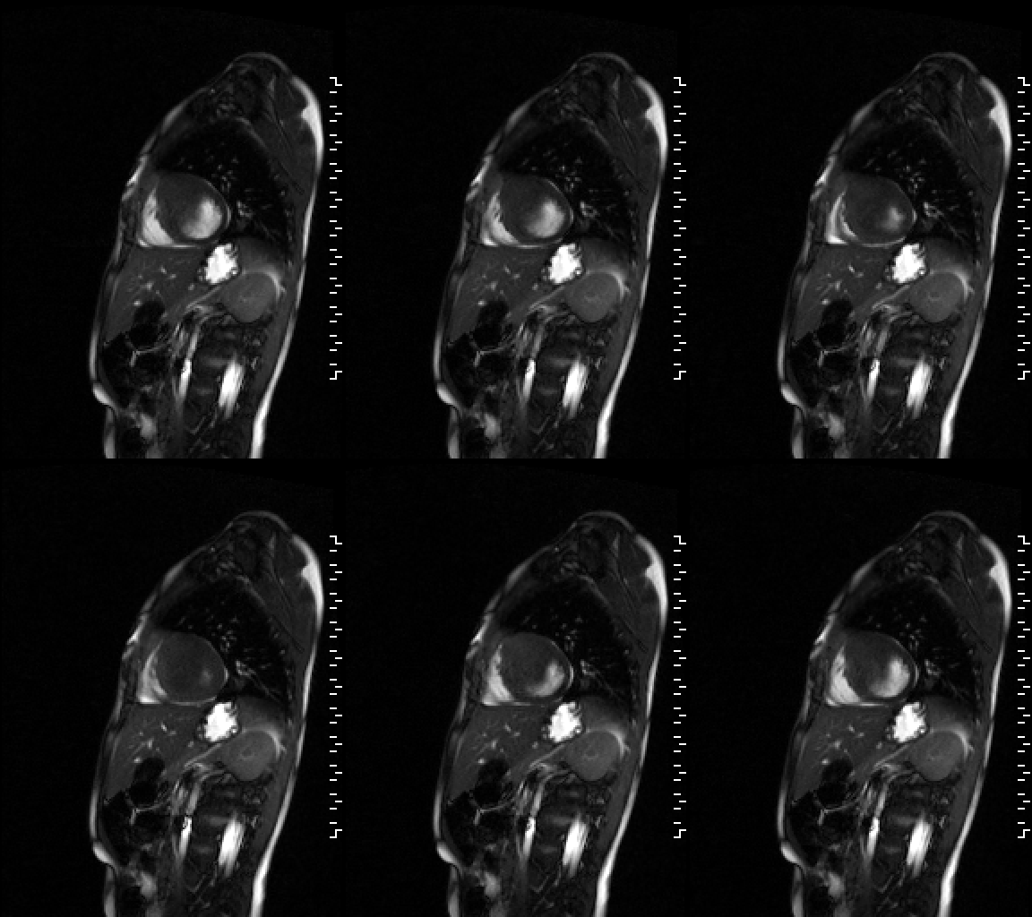
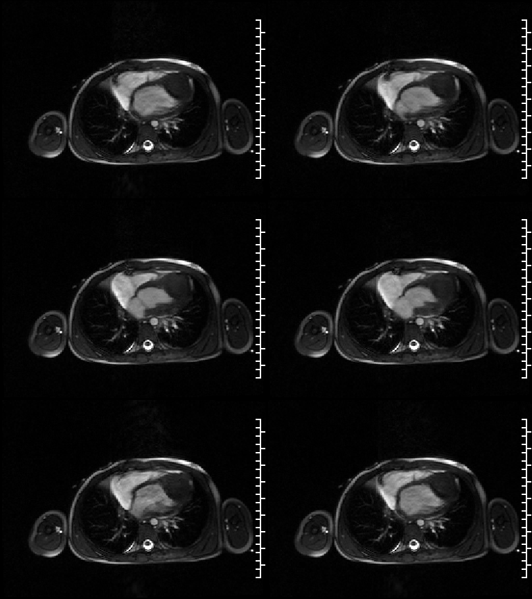
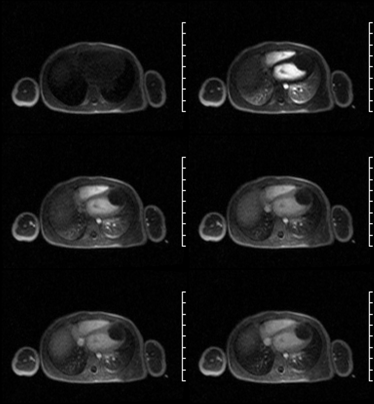
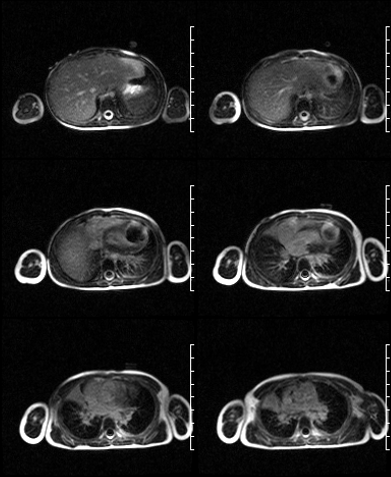
Radiologic Findings Selected Cardiac MRI images. GRE sagittal (Fig. 1) and GRE 4-chamber view (Fig. 2) cine images demonstrate a large, 3.0 cm x 3.0 cm x 4.0 cm, well circumscribed, soft-tissue mass involving the left ventricle. The epicenter of the mass is localized to the anteroseptal and septal wall with sparing of the extreme cardiac apex and base. The mass extends into the anterior wall and a portion of the anterolateral wall, encroaching on the ventricular cavity itself. First pass axial image during the infusion of Gd-DTPA (Fig. 3) reveals no tumor enhancement. 10 minute delayed post-contrast (Gd-DTPA) axial SSFP sequence reveals heterogeneous predominantly peripheral enhancement of the mass (Fig. 4). No signal dropout from calcium in appreciated within the mass on any of the provided pulse sequences. Answer Diagnosis: Cardiac Fibroma
Answer
Diagnosis: Cardiac Fibroma
Differential Diagnosis
Cardiac Rhabdomyoma
Discussion
Cardiac fibroma is a rare, albeit benign, tumor of the heart. Synonyms include cardiac fibromatosis, fibrous hamartoma, and fibroelastic hamartoma. Most cases present in infancy or early childhood. In fact, cardiac fibroma is the second most common benign primary cardiac tumor in children after rhabdomyoma. Grossly, cardiac fibroma ranges 2-10 cm in diameter with a mean of 5.0 cm. Fibromas usually have no foci of cystic degeneration, hemorrhage, or necrosis. Dystrophic calcification is however, common. Histologically, the tumor is composed of fibroblasts and collagen. The tumor is most often located in the ventricular septum or the ventricular wall. One third of patients are asymptomatic. Symptoms depend on the size of the tumor. Larger tumors involving the conduction system of the heart may present with cardiac dysrhythmias Tumors obstructing blood flow may present with cardiac decompensation. An increased association with Gorlin syndrome has been described.
Cardiac rhabdomyoma Cardiac rhabdomyoma is the most common benign primary cardiac tumor in children. It is composed of striated muscle tissue. This tumor is closely associated with tuberous sclerosis and occurs in more than 50% of patients with tuberous sclerosis. Although rhabdomyoma may affect any chamber of the heart, it most frequently affects the left ventricle. This tumor may present as a single lesion or as multiple, non-capsulated soft lesions. Most patients are asymptomatic. Clinical presentations include heart failure secondary to outflow obstruction, cardiac dysrhythmias, embolic disease and sudden death.
Imaging features
Fibroma
MRI
- Isointense relative to muscle on T1WI
- Usually homogeneous and hypointense on T2WI
- No contrast uptake during perfusion imaging
- May demonstrate delayed enhancement; often in a heterogeneous fashion from periphery to center
Rhabdomyoma
MRI
- Isointense relative to muscle on T1WI
- Increased signal intensity on T2WI
- Hypointense to myocardium after contrast administration
Suggested Readings
- Araoz PA, Mulvagh SL, Tazelaar HD, et al. CT and MR Imaging Features of Benign Primary Cardiac Neoplasms with Echocardiographic Correlation. RadioGraphics 2000; 20: 1303-1319.
- Sparrow PJ, Kurian JB, Jones TR, Sivananthan MU. MR imaging of Cardiac Tumors. RadioGraphics 2005; 25: 1255-1276.
Original case written by its authors at Virginia Commonwealth University and published at this address as part of a weekly teaching collection. Reproduced here as an archive.